Treatment of Hip Osteoarthritis (Coxarthrosis)
Hip osteoarthritis is gradual wear of hip cartilage, typically causing groin pain and limping. This page explains causes, contributing conditions such as dysplasia and childhood hip dislocation, non-surgical approaches and how replacement decisions are made.
Also known as: Coxarthrosis, Coxarthrosis, Hip osteoarthritis, Hip osteoarthritis
Your Treatment Pathway
Examination and X-ray
The source of groin pain, movement restriction and standing pelvic X-rays establish the stage.
Plan
Non-surgical or surgical care is selected according to stage, age, dysplasia or dislocation history and expectations.
Administration
Weight management and exercise, medication and image-guided injection when needed; replacement planning in advanced disease.
Review
Pain, walking distance and X-rays are reassessed at intervals.
Movement
Goal: pain control, preserving hip movement and independent walking.
What Is Hip Osteoarthritis?
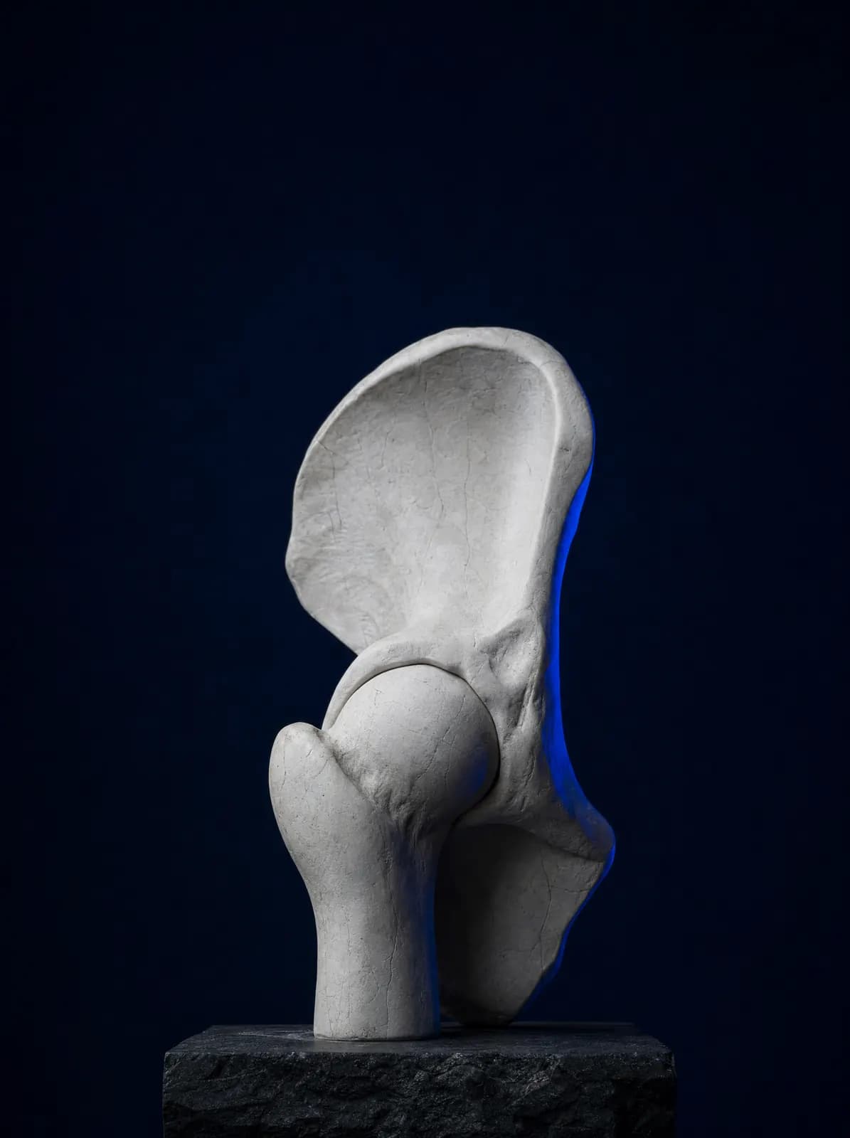
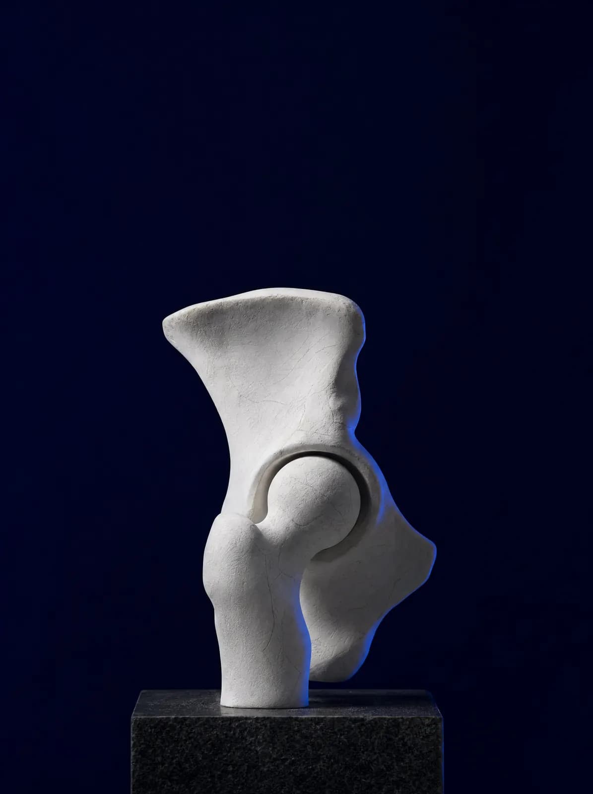
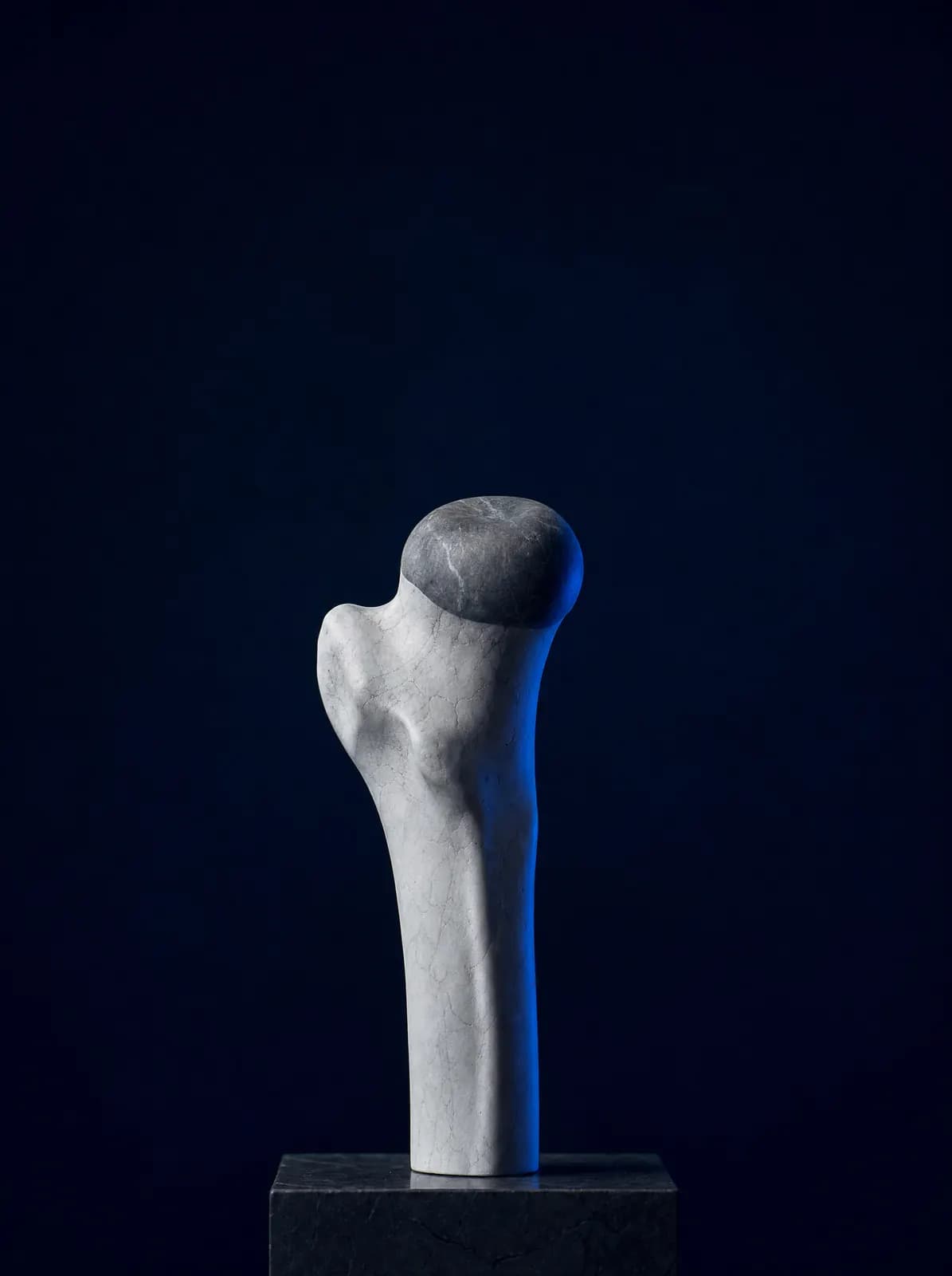
The hip is a ball-and-socket joint formed by the rounded end of the thigh bone (femoral head) and the socket in the pelvis (acetabulum). Both surfaces are covered with cartilage, which distributes body weight during walking and allows smooth movement. Osteoarthritis involves thinning and deterioration of this cartilage, with the bone surfaces gradually moving closer together.
Although commonly called "calcification" in Turkish, osteoarthritis does not involve lime accumulating in the joint; the name comes from the bony spurs seen on X-rays. Hip osteoarthritis usually develops slowly over years. Common contributing factors include excess weight, heavy lifting at work, previous hip fracture or dislocation, impaired femoral head blood supply and a shallow socket present from birth (dysplasia).
Not all hip pain is osteoarthritis. Hip impingement causing a pinching sensation in the groin, tendon or bursal problems causing outer hip pain, and nerve compression in the lower back can produce similar symptoms. Examination and X-rays distinguish them; an overview is available on the hip conditions page.
What Are the Symptoms?
One surprising feature of hip osteoarthritis is that pain is often felt in the groin, not behind the hip. It may spread to the front-inner thigh or even the knee, so it can initially be mistaken for a knee problem.
- Pain in the groin, inner thigh or side of the hip that increases with walking
- Stiffness lasting 10–20 minutes after waking or prolonged sitting
- Difficulty tying shoelaces, putting on socks and getting into a car
- Reduced walking distance and limping
- Restricted movement when moving the leg to the side or rotating it inwards and outwards
- Night pain and a feeling of leg shortening in advanced disease
Hip Osteoarthritis in Older People
In older people, the aim is to preserve independent walking and safe balance. Reduced movement due to pain weakens muscles and increases fall risk, so exercise is adapted rather than stopped. Accompanying osteoporosis is addressed together; see orthopaedics in older age and osteoporosis.
How Is It Diagnosed?
Diagnosis rests on examination and a standing pelvic X-ray. X-rays show narrowing of the joint space, bony spurs, hardening beneath the cartilage and cysts; socket depth and differences in leg length are also measured. Pain and restricted hip rotation during examination largely establish the diagnosis.
MRI is not needed for every patient. It is requested when pain persists despite near-normal X-rays, avascular necrosis is suspected or a labral or cartilage problem needs investigation. Blood tests do not diagnose osteoarthritis; they help distinguish inflammatory rheumatic disease. The lower back is also examined if pain may originate there.
How Does Hip Dysplasia Lead to Osteoarthritis in Adults?
Acetabular dysplasia means the hip socket has not developed sufficient depth. A shallow socket concentrates body weight on a narrow edge rather than a broad area. This focal loading wears the cartilage and rim tissue (labrum) prematurely over the years. Osteoarthritis may therefore develop younger in people with dysplasia than in the general population.
Symptoms often begin in the 20s–40s as groin pain after prolonged walking or standing. Diagnosis is made by measuring socket angles on a standing pelvic X-ray. Early management includes muscle strengthening, weight control and reducing load; when the socket is markedly shallow but cartilage is still preserved, bone surgery to reorient the socket (acetabular osteotomy) may be considered. If much of the cartilage has been lost, management follows the treatment pathway for osteoarthritis.
Screening and early treatment in infancy are the most important preventive steps. Developmental hip dysplasia in babies is a separate topic, explained on the developmental dysplasia of the hip in babies page.
What Problems May Develop Later in People Who Had a Dislocated Hip in Childhood?
In people treated for hip dislocation during infancy or childhood, the joint may not function exactly like a normal hip throughout life. Residual mismatch between the socket and femoral head distributes cartilage loading unevenly; early osteoarthritis, leg-length difference, limping and muscle fatigue may develop in adulthood. The effects are more pronounced when childhood dislocation was untreated.
Treatment is individualised: shoe lifts for leg-length difference, targeted exercises for muscle balance, non-surgical pain management and replacement in advanced cases are discussed. An abnormal socket makes replacement technically more complex to plan; this is explained clearly at examination. General information is available on the hip replacement surgery and recovery page.
What Are the Non-Surgical Treatment Options?
Three foundations of non-surgical care apply at every stage: weight control, muscle strengthening and range-of-movement exercises, particularly for the muscles at the side of the hip, and activity modification. Painkillers, anti-inflammatory medicines, physiotherapy and a walking stick held opposite the painful side may be added. A stick substantially reduces hip loading and is nothing to be embarrassed about.
Because the hip joint lies deep within the body, intra-articular injections are less straightforward than in the knee and are usually performed with imaging guidance. The effects of hyaluronic acid, PRP and corticosteroid injections in hip osteoarthritis vary between patients. Claims that regenerative methods such as stem cells and exosomes renew cartilage remain under investigation, and these treatments are not suitable for every patient; the methods are compared on the regenerative and non-surgical treatments page.
When Is Surgery Needed?
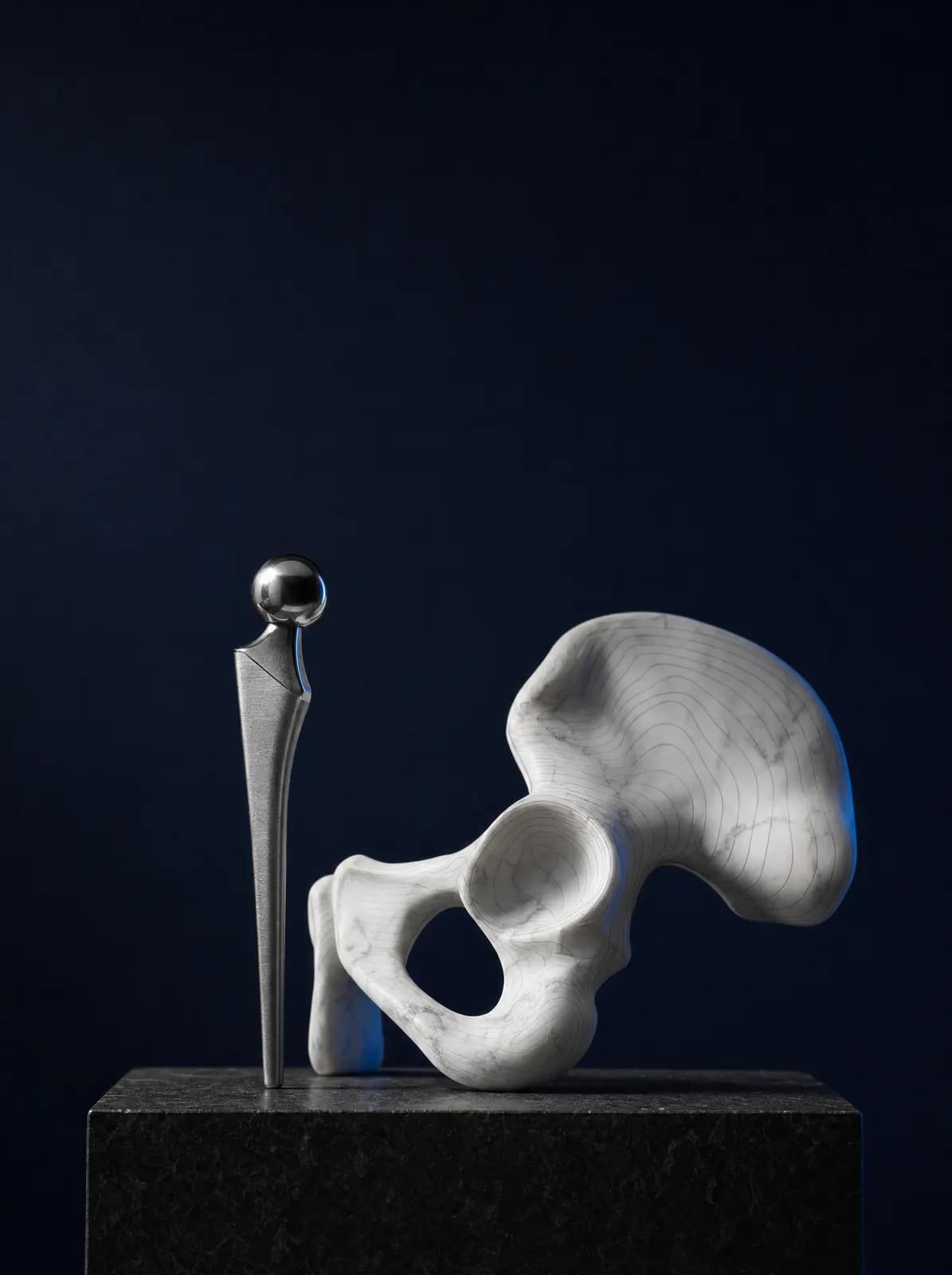
Surgery is not decided from X-rays alone. Hip replacement is discussed when pain interrupts sleep despite non-surgical treatment, walking distance is substantially reduced, everyday tasks such as putting on shoes become difficult, and X-rays show severe joint-space narrowing.
In younger patients with dysplasia and largely preserved cartilage, bone surgery to reorient the socket may be discussed instead of joint replacement. The decision is made together, considering age, weight, occupation, expectations and coexisting conditions. The procedure, hospital stay, early walking and movements requiring care are explained on the hip replacement and recovery page.
What Should You Consider in Daily Life?
Walking on level ground, cycling and especially swimming preserve muscle strength without overloading the hip. Prolonged squatting, sitting cross-legged and using low chairs or toilets increase hip loading; raising seat height may reduce pain. Dividing loads instead of lifting heavy objects and taking breaks during longer walks are helpful.
Weight management is one of the most effective measures for hip osteoarthritis. During walking, the hip bears several times body weight, so even modest weight loss can affect pain. Consistent exercise is part of treatment: it should be regarded as a daily habit rather than a programme lasting a few weeks. At AC CLINIC, programmes for patients from Ordu and the surrounding area are adapted to their work and walking habits.
Frequently Asked Questions
The hip joint lies deep in the body behind the groin. Its nerves supply the groin and front-inner thigh, so pain is commonly felt there and sometimes radiates to the knee. Some pain thought to arise from the knee therefore originates in the hip.
Worn cartilage does not return to its original state spontaneously. Weight control, suitable exercise and load management can reduce symptoms and slow progression; results vary and are discussed realistically at consultation.
Consult an orthopaedics and traumatology specialist for diagnosis and treatment planning. Exercise may be coordinated with physiotherapy when needed.
They may be related. A hip dislocation treated or missed in childhood can leave imperfect joint congruity and lead to early adult osteoarthritis. A standing pelvic X-ray assesses this.
No. Regular walking on level ground, cycling and swimming are recommended. Prolonged squatting, cross-legged sitting, low seating and carrying heavy loads are more demanding.
You can book by telephone or WhatsApp at AC CLINIC in Altınordu. Bringing previous pelvic X-rays and MRI scans helps the assessment.
References
Appointments and Contact
Call or contact us on WhatsApp to arrange an appointment. Briefly describe your symptoms; we try to schedule your examination and any necessary imaging on the same day.
- Opening hours
- 08:30–18:00Monday – Saturday
The clinic is not a 24/7 emergency department. For a fracture, dislocation, open wound, or inability to bear weight with fever, call 112 or attend the nearest hospital emergency department.