Avascular Necrosis of the Femoral Head
Avascular necrosis of the femoral head is death of bone tissue caused by impaired blood flow to the rounded upper end of the femur in the hip. Joint-preserving options exist when diagnosed early; as it progresses, the head collapses and hip replacement may be considered.
Also known as: Avascular necrosis (AVN), Osteonecrosis, Femoral head necrosis, Hip osteoporosis and osteonecrosis

Your Treatment Pathway
Examination and Imaging
The source of groin pain is investigated; MRI is requested if suspected even with normal X-rays.
Staging
The extent of the necrotic area and whether the femoral head has collapsed are assessed.
Plan
Joint-preserving options before collapse and a replacement plan after collapse are discussed.
Treatment and Follow-up
Reducing load, addressing risk factors and arranging follow-up imaging at intervals.
Movement
Aim: pain control, preservation of the joint where possible and continued walking.
What Is Avascular Necrosis of the Femoral Head?
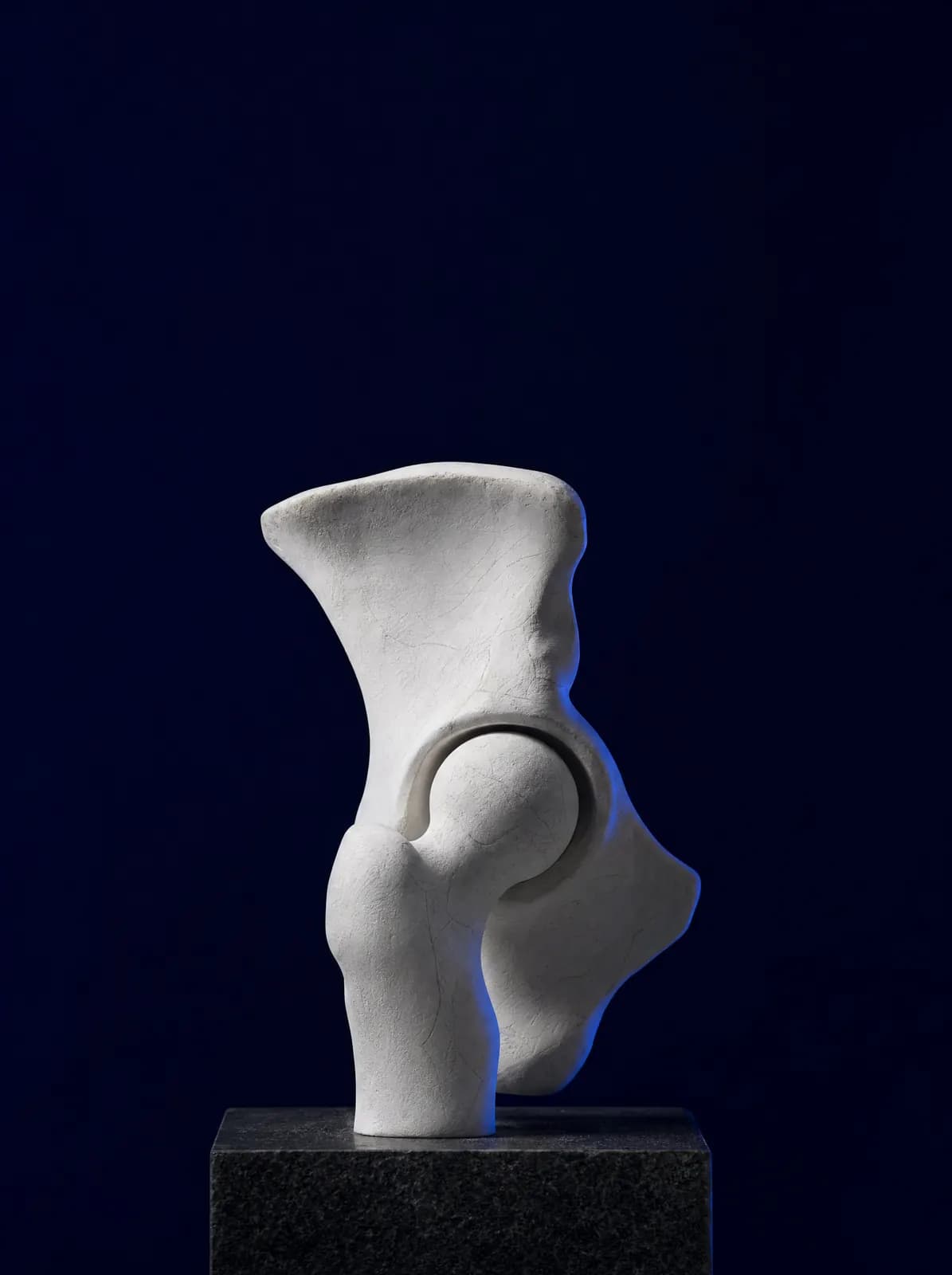
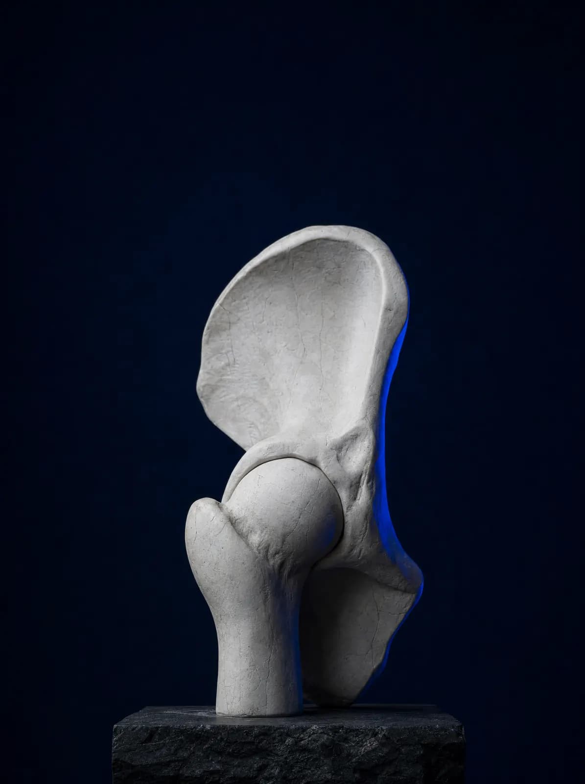
The rounded end of the thigh bone within the hip joint is called the femoral head. It is supplied by small blood vessels and has limited alternative circulation. If blood flow falls for any reason, bone cells lose their viability; this is called avascular necrosis (osteonecrosis), commonly described as 'decay' or 'loss' of the hip bone.
Dead bone initially retains its shape. Over time it weakens and the surface collapses under load; once the spherical shape is lost, cartilage rapidly wears and hip osteoarthritis develops. The crucial point is therefore diagnosis before collapse.
Avascular necrosis can occur at any age but is most common at 30–50, and both hips are affected in many patients. The other hip is therefore assessed when one side is diagnosed.
What Are the Causes and Risk Factors?
No clear cause is found in some patients. When one is identified, common causes include:
- Long-term or high-dose cortisone (steroid) use
- Regular heavy alcohol consumption
- Previous fracture or dislocation around the hip (post-traumatic impairment of blood supply)
- Sickle cell anaemia and certain blood disorders
- Decompression sickness seen in divers (the bends)
- Rheumatic diseases and the medicines used to treat them
- Radiotherapy and certain chemotherapy medicines
Post-Traumatic Necrosis
Femoral neck fractures and hip dislocations can directly damage vessels supplying the femoral head. Necrosis may therefore develop months or years after a hip fracture even when it has united, which is why follow-up continues. For the fracture itself, see hip fractures in older people.
What Are the Symptoms?
Early disease may be symptom-free and discovered on imaging performed for another reason. When symptoms start, deep groin pain that increases with walking is typical.
- Throbbing groin pain that may spread to the front of the thigh and knee
- Pain increasing with walking and standing and partly relieved by rest
- At a later stage, night pain and discomfort that persists at rest
- Limping and reduced walking distance
- Marked restriction of hip movement after femoral head collapse
How Is It Diagnosed?
The first steps are examination and a standing pelvic X-ray. However, early X-rays may look normal, which does not exclude disease. If suspicion remains, MRI is requested. It can show non-viable bone before the shape changes, making it central to early diagnosis.
The disease is staged according to MRI findings and whether X-rays show collapse. Broadly, pre-collapse stages, in which the head retains its spherical shape, are distinguished from post-collapse stages, in which the surface has collapsed and cartilage is damaged. Treatment planning depends directly on this distinction. Both hips are assessed even if only one hurts because bilateral involvement is common.
What Are the Non-Surgical Treatment Options?
In the early stage, the aim is to reduce load on the bone and allow time for healing. Basic measures include using crutches to reduce weight-bearing, painkillers and anti-inflammatory medicines, weight control and avoiding strenuous activity. Any contributing factor such as cortisone or alcohol is reviewed with the relevant doctor.
Certain medicines affecting bone formation and supportive treatments may be considered; their effect on disease progression varies between individuals and does not carry a promise of a definite result. The role of regenerative procedures such as stem cells, PRP and exosomes in avascular necrosis remains under investigation: although some studies report promising results in early disease, evidence is limited and these procedures are not suitable for everyone. How these methods are used and the context in which they are discussed are explained on the stem cell treatment page.
Non-surgical observation alone may be insufficient for large areas of necrosis with a high risk of collapse; follow-up intervals and decision points are therefore clearly established during consultation.
When Is Surgery Needed?
Surgical options fall into two groups. The first comprises joint-preserving operations, which are meaningful before collapse: core decompression, which creates a channel in the bone to reduce pressure and support blood supply; additional bone grafting; and, in selected patients, corrective osteotomies that shift loading onto healthy bone.
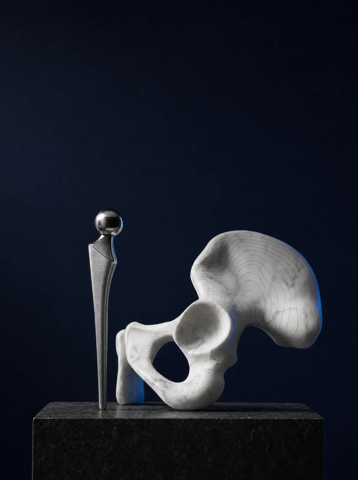
The second group is hip replacement, considered after the bone head has collapsed and the cartilage has deteriorated. At this stage, advanced joint damage is the source of pain, and treatment aims to reduce pain and restore walking capacity. The operation, recovery timetable and precautions are covered in hip replacement surgery and recovery.
The pathway is agreed together according to age, the extent of necrosis, whether collapse has occurred and patient expectations. No method's result can be predicted with certainty; potential benefits and risks are discussed individually during assessment.
What Should You Consider in Daily Life?
After diagnosis, regular follow-up is essential: do not miss imaging reviews even if symptoms improve, because collapse can begin silently. Using crutches to reduce loading for the recommended period, avoiding impact activities such as jumping and running, and controlling weight help protect the joint.
Stopping alcohol consumption and using corticosteroid-containing medicines only on medical advice are modifiable factors that may affect progression. Low-impact activities such as swimming and cycling are generally recommended. At AC CLINIC, follow-up intervals for patients from Ordu and the surrounding area are planned according to the stage of necrosis and occupational loading.
Frequently Asked Questions
Small areas of necrosis outside weight-bearing regions may remain silent, but larger areas tend towards collapse over time. Regular imaging therefore continues even if symptoms improve.
Yes. Early X-rays may look normal. Persistent suspicion warrants MRI, which can show non-viable bone before its shape changes.
The role of stem cells and similar regenerative treatments in this condition remains under investigation. Some early-stage studies report promising results, but evidence is limited and the method is not suitable for everyone; suitability is assessed during consultation.
Both sides are often affected. When one hip is diagnosed, the other is also imaged and monitored even if painless.
Do not stop the medicine yourself. Cortisone is often essential for another condition; dose and duration must be reviewed with the prescribing doctor.
You can book by phone or WhatsApp at AC CLINIC in Altınordu. Bringing MRI and X-ray images on CD or as files helps staging.
References

Appointments and Contact
Call or contact us on WhatsApp to arrange an appointment. Briefly describe your symptoms; we try to schedule your examination and any necessary imaging on the same day.
- Opening hours
- 08:30–18:00Monday – Saturday
The clinic is not a 24/7 emergency department. For a fracture, dislocation, open wound, or inability to bear weight with fever, call 112 or attend the nearest hospital emergency department.