Bone and Soft-Tissue Tumours: Diagnosis, Biopsy and Surgery (Orthopaedic Oncology)
A swelling in an arm, leg or the trunk, bone pain worsening at night, or a bone lesion on imaging should be assessed in the correct sequence. This page outlines the next steps according to your symptoms.
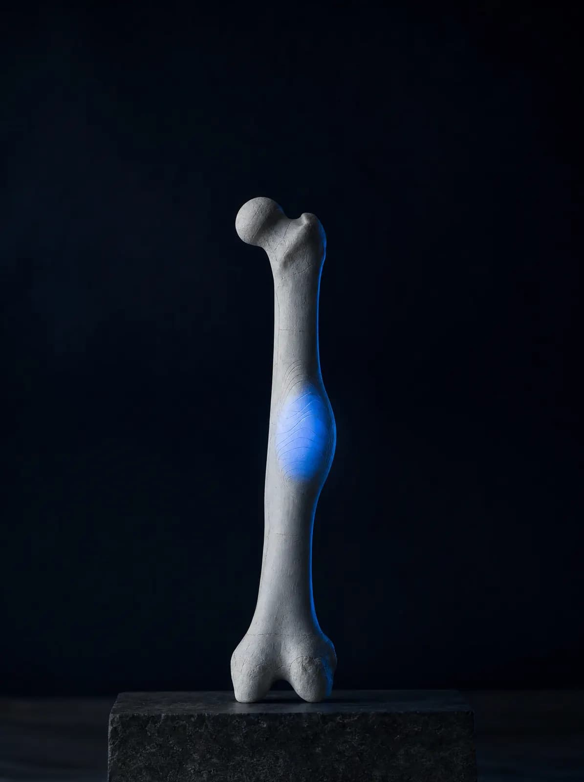
Diagnosis and treatment of masses in bone and soft tissue (muscle, fat and connective tissue) fall within orthopaedic oncology. The most important rule in this field is the sequence: history and examination first, then appropriate imaging, a correctly performed biopsy if needed, and treatment last. Disrupting this sequence, for example by removing a mass without prior planning, can make subsequent treatment more difficult. Initial assessment of suspicious masses by a team familiar with these principles is therefore recommended.
Most palpable lumps and bone lesions seen on scans are benign. Some need no treatment and are simply monitored at intervals. Others are surgically cleared because they weaken the bone or cause pain. Malignant tumours are uncommon, but prompt assessment matters because early diagnosis directly affects treatment planning and the chance of preserving the limb.
Patients most often ask, "Is my lump dangerous?" The answer depends on the size, growth rate, depth, pain pattern, age and imaging findings together, not on a single sign. Being painless does not automatically make a lump harmless; rapidly growing lumps, those larger than 5 cm and those beneath the muscle layer are assessed more closely.
Prof. Dr. Alper Çıraklı is a member of the TOTBİD Bone and Soft Tissue Tumours Branch; assessment of lumps, imaging plans and biopsy decisions follow these principles. If a malignancy is diagnosed, treatment is planned jointly with medical and radiation oncology teams. The table below links common descriptions used by patients to the relevant section. You can book by phone or WhatsApp for an examination and review of your existing images at AC CLINIC in Altınordu, Ordu.
By Symptom
Which Swellings and Pains Should Be Assessed?
Not every lump is a tumour; cysts, fatty lumps, inflammatory swelling, collections after muscle tears and thickening after old injuries can look similar. Nevertheless, some features require detailed investigation. Do not postpone assessment if any of the following are present:
- Swelling that grows noticeably over weeks or months
- A firm, poorly mobile mass beneath the muscle layer
- A soft tissue mass larger than approximately 5 cm
- Bone pain that worsens at night, persists at rest and responds poorly to painkillers
- A fracture after minor strain (suspected pathological fracture)
- Increased blood vessels over the mass, changes in skin colour or temperature
- Swelling with unexplained weight loss, fever or night sweats
Does a Painless Mass Mean There Is No Cause for Concern?
No. Some soft tissue lumps grow without pain; pain may start only when they compress a nerve or blood vessel. Decisions therefore depend on size, depth and growth rate rather than pain. A growing lump needs imaging even if painless.
When to Seek Care Without Delay
Do not wait if a fracture occurs without a clear accident, the limb cannot bear weight, a tense swelling grows rapidly, or redness and warmth accompany fever. The clinic is not a 7/24 emergency department; call 112 or attend the nearest hospital emergency department.
Does a Mass Always Arise from Bone?
No. Soft-tissue masses arising from muscle, fat and connective tissue are also common, and the great majority are benign (most commonly lipomas). However, deep, firm masses larger than approximately 5 cm that continue to grow require detailed investigation; they should not be removed without planning. A soft-tissue mass noticed in Ordu is assessed for size, depth and growth rate during consultation; a detailed page is being prepared.
How Is a Biopsy (Tissue Sampling) Performed? Open and Closed Biopsy
Biopsy takes a small sample from a mass for microscopic examination. Imaging shows its location and behaviour, but pathology often provides the definitive diagnosis. Because treatment planning depends on that result, biopsy is the first step in treatment.
A closed (needle) biopsy uses a special needle passed through the skin, with ultrasound or CT guidance if required. Local anaesthesia is usually sufficient, there is no incision scar and the patient goes home the same day. An open biopsy obtains tissue directly through a small incision; it is preferred when a needle biopsy does not provide enough material or a larger sample is needed for diagnosis.
The site and direction of a biopsy affect the planning of any later operation: the biopsy tract is chosen to lie within the area that would be removed during subsequent surgery. It is therefore recommended that the biopsy be planned by, or in collaboration with, the team that will operate. Concern that a biopsy 'awakens or spreads the tumour' is common; this is not an expected outcome of a correctly performed biopsy, whereas starting treatment without a diagnosis poses a real risk.
How Does Care Progress from Diagnosis to Treatment?
The process is staged. After examination and initial imaging, MRI, CT or a bone scan may be requested according to the lump's features. A biopsy is performed if needed. Pathology results guide monitoring, surgery or multidisciplinary treatment; these options are detailed on the bone tumours page.
Decisions about malignant tumours are not made by one doctor alone, but through collaboration between orthopaedics, medical oncology, radiation oncology, radiology and pathology. Prof. Dr. Alper Çıraklı's membership of the TOTBİD Bone and Soft Tissue Tumours Branch is part of his involvement in this field. Assessment in Ordu involves evaluating symptoms in the correct sequence, arranging imaging and biopsy, and coordinating care with a specialist treatment centre when needed.
Patients often ask how long the process will take. Imaging and biopsy are generally completed within days; pathology results may take a few working days to several weeks if additional stains are needed. Waiting is difficult, but starting the correct treatment depends on this result.
Other Conditions Mistaken for a Mass: Foreign Bodies, Infection and Deformity
Not every swelling is a tumour: a retained foreign body may cause a firm area, bone or joint infection can mimic a mass with swelling, warmth and pain, and an old healed fracture or congenital deformity may be felt as a bony prominence. These subjects have their own pages on the site, linked here.
Other Topics
The topics below are also commonly encountered in this area. Detailed pages are being prepared; matching symptoms can be assessed at consultation.
- Soft tissue tumours (lipoma, lumps, sarcoma): when to be concerned and how surgery is planned
- Mole (naevus) excision: removal of suspicious or troublesome moles and submission for pathology
- Callus excision: surgical treatment of deep plantar calluses that impede walking
- Cryotherapy (freezing) for wart treatment
- A monitoring programme for bone cysts and incidentally detected lesions
Frequently Asked Questions
Initial assessment is by an orthopaedics and traumatology specialist; a doctor with an interest in bone and soft tissue tumours (orthopaedic oncology) can streamline the process. If malignancy is diagnosed, treatment is planned with medical and radiation oncology.
X-ray is usually the first test for bone symptoms, showing bone structure and lesion boundaries. Ultrasound may be the first step for soft-tissue masses, with MRI for detailed assessment. CT and bone scintigraphy are added selectively. The necessary investigations are decided after examination.
A biopsy performed with the correct technique and at the appropriate site is not expected to spread the tumour. The main risk is an unplanned procedure before diagnosis, which can complicate the margins of later surgery. Biopsy should therefore be planned with the team that will provide treatment.
No. Many masses that do not grow or cause symptoms and appear benign on imaging need only periodic monitoring. Surgery is considered for reasons such as growth, pain, bone weakening, loss of function or the need to clarify the diagnosis.
Most results are available within a few working days. Additional staining or molecular testing may extend this to several weeks. Waiting is necessary to choose the correct treatment; once results arrive, the plan is explained fully during consultation.
You can book by phone or WhatsApp at AC CLINIC in Altınordu. Bringing previous X-rays, ultrasound, MRI or CT images on film or CD, any pathology reports and a medication list helps the assessment.
Appointments and Contact
Call or contact us on WhatsApp to arrange an appointment. Briefly describe your symptoms; we try to schedule your examination and any necessary imaging on the same day.
- Opening hours
- 08:30–18:00Monday – Saturday
The clinic is not a 24/7 emergency department. For a fracture, dislocation, open wound, or inability to bear weight with fever, call 112 or attend the nearest hospital emergency department.