Knee Pain and Knee Conditions: Treatment
Knee pain can have many causes, from osteoarthritis and meniscal tears to ligament injuries and fractures. This page guides you to the relevant section according to your symptoms.
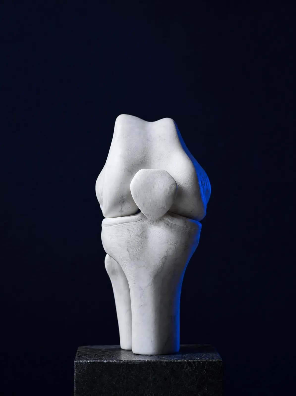
The knee is one of the body's most heavily loaded and frequently moving joints. The thigh bone (femur), shin bone (tibia) and kneecap (patella) meet here; their surfaces are covered with cartilage, the menisci between them act as shock absorbers, and the cruciate and collateral ligaments hold the joint together. When any of these structures is strained, the resulting symptom is often the same: knee pain. Distinguishing the cause requires a history, examination and, when necessary, imaging.
How symptoms begin often provides clues. Pain that gradually increases over years and is worse when descending stairs or squatting is often associated with osteoarthritis. Catching and locking after a sudden twist suggest a meniscal tear, while a "pop" during sport followed by swelling and a feeling of giving way suggests a ligament injury. Inability to bear weight, deformity or rapidly increasing swelling after a fall or blow requires urgent assessment.
Treatment decisions are not based on imaging findings alone. Age, occupation, sporting habits, leg alignment, weight and coexisting conditions directly affect the plan. Two patients with identical X-rays may be offered different approaches; the reasons are explained clearly during the consultation. For a substantial proportion of knee problems, first-line treatment is non-surgical: activity modification, an exercise programme targeting the quadriceps, weight management, medication and, when needed, intra-articular treatments. Surgery is considered when these measures are insufficient or when a structure needs repair.
The table below links patients' most common descriptions to the relevant page. If your symptoms fit several rows, start with the most troublesome; the whole picture is assessed together during examination. You can book by telephone or WhatsApp at AC CLINIC in Altınordu, Ordu, for knee examination, planning of necessary imaging and discussion of treatment options.
By Symptom
Topics
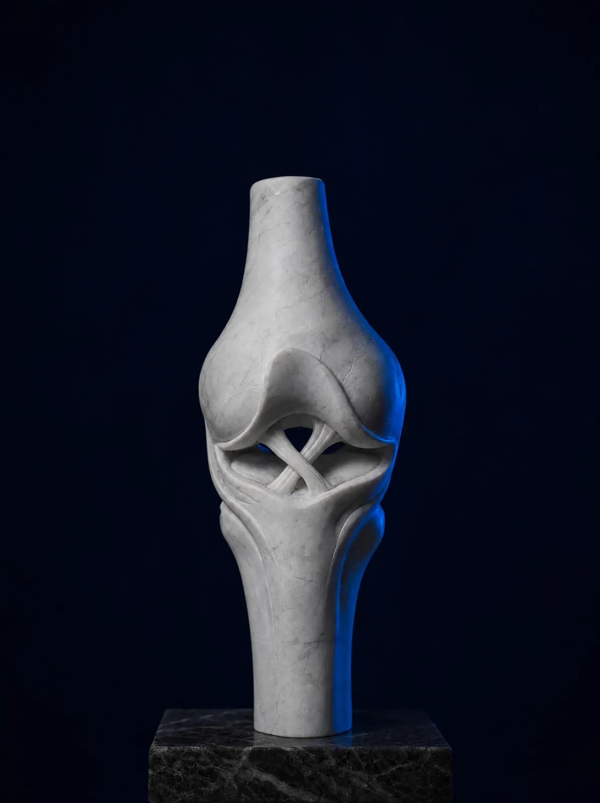

What Are Posterior Cruciate Ligament (PCL) and Multiple Ligament Injuries?
The posterior cruciate ligament (PCL) is the strong ligament that prevents the shinbone sliding backwards relative to the thighbone. It is injured much less often than the anterior cruciate ligament, usually during a high-energy event: a heavy blow to the front of a bent knee (striking a dashboard or falling onto the knee during football). Patients usually do not describe a 'pop'; instead, they report fullness behind the knee, difficulty squatting and walking downhill, and instability on stairs.
Many isolated partial PCL injuries are managed without surgery: swelling control, progressive quadriceps strengthening and a specialist support brace when needed. Surgery is discussed for complete rupture, persistent marked instability or injury to several ligaments together.
Multiligament injury (rupture of more than one ligament at the same time) is a separate condition: it may accompany knee dislocation, carries a risk of vessel and nerve injury, and requires urgent assessment. Circulation checks, appropriate stabilisation and surgical planning are prioritised; it should not be confused with an isolated anterior cruciate ligament injury.
What Is Knee Bursitis, and How Is It Treated?
A bursa is a thin fluid-filled sac that reduces friction between bone and skin or tendon. Irritation of the bursae at the front of the knee, above and below the kneecap, is called bursitis. It occurs in people who spend long periods kneeling, such as carpet or flooring fitters, cleaners and agricultural workers, after repeated knocks, and sometimes without trauma. Symptoms include a soft, well-defined swelling at the front of the knee, tenderness that increases with pressure and difficulty kneeling.
Bursitis and fluid accumulation inside the joint are different: bursitis lies just beneath the skin, whereas joint fluid causes swelling of the knee as a whole and restricts movement. Examination distinguishes between them. Treatment usually consists of stopping the triggering activity, applying cold, using a knee support or kneeling protection, taking anti-inflammatory medication and, when needed, draining the fluid. Redness, warmth and fever over the swelling require prompt assessment because of possible infection; do not attempt to puncture or drain it yourself.
Fractures Around the Knee: Patella and Tibial Plateau
A fall, road traffic accident or landing from a height can fracture the kneecap (patella), lower end of the thighbone or upper surface of the shinbone (tibial plateau). Inability to bear weight, rapidly increasing swelling, deformity and inability to lift the leg straight are typical warning signs. Some of these fractures involve the joint surface and require detailed imaging for planning; general fracture treatment, fixation and recovery are discussed on the relevant page.
Foreign Bodies, Masses, Infection and Deformity in the Knee
Foreign bodies in tissues around the knee, growing masses, bone and joint infections, and bow legs or knock knees each have distinct assessment pathways. These subjects are explained in detail on their own pages, linked here; follow the one relevant to your symptoms.
Other Knee Topics
The following are also common knee problems seen in consultation. Detailed pages are being prepared; symptoms matching any of these subjects are assessed fully during examination.
- Discoid meniscus (a congenitally different meniscal shape)
- Recurrent kneecap dislocation (patellar instability)
- Knee cartilage damage and osteochondritis dissecans
- Loose body (a piece of cartilage or bone moving freely within the joint)
Frequently Asked Questions
An orthopaedics and traumatology specialist assesses knee pain and plans treatment. Exercise and rehabilitation may be coordinated with physiotherapy; rheumatology advice may be requested if rheumatic disease is suspected.
The first step is generally a standing knee X-ray to show joint space, bone structure and leg alignment. MRI is not required for every patient. It is requested if meniscal, ligament or cartilage injury is suspected or X-rays do not explain the findings.
Inability to bear weight, deformity after a fall or accident, rapidly increasing swelling, knee redness and warmth with fever, or foot numbness or colour change are emergency signs. The clinic is not a 7/24 emergency service; call 112 or attend the nearest hospital emergency department.
No. Most meniscal and ligament operations are arthroscopic, whereas joint replacement, alignment correction and fracture fixation are open operations. Suitability depends on the diagnosis, tissue condition and patient expectations.
A knee brace is not a treatment alone but offers support in selected situations. Protective braces after ligament injury and specialised unloading braces for osteoarthritis may help. Continuous unnecessary use may weaken the thigh muscles, so the decision should follow examination.
You can book by telephone or WhatsApp at AC CLINIC in Altınordu. Previous X-ray, MRI or CT images and a list of your medicines help assessment.
Appointments and Contact
Call or contact us on WhatsApp to arrange an appointment. Briefly describe your symptoms; we try to schedule your examination and any necessary imaging on the same day.
- Opening hours
- 08:30–18:00Monday – Saturday
The clinic is not a 24/7 emergency department. For a fracture, dislocation, open wound, or inability to bear weight with fever, call 112 or attend the nearest hospital emergency department.