Knee Osteoarthritis (Gonarthrosis) Treatment
Knee osteoarthritis is wear of the knee cartilage over years. Early stages are managed with exercise, weight control and injections; knee replacement is considered in advanced disease. This page explains which options are discussed at each stage and why.
Also known as: Gonarthrosis, Knee osteoarthritis, Knee osteoarthritis, Osteoarthritis of the knee joint
Your Treatment Pathway
Examination and X-ray
Pain location, range of movement and weight-bearing X-rays determine the stage.
Plan
The non-surgical or surgical approach is chosen together according to the stage, age and expectations.
Administration
An exercise and weight management programme, injections if necessary, and joint replacement planning at an advanced stage.
Review
Monitoring of pain, walking distance and X-rays at regular intervals.
Movement
Aim: controlling pain in daily life and preserving walking distance.
What Is Knee Osteoarthritis?
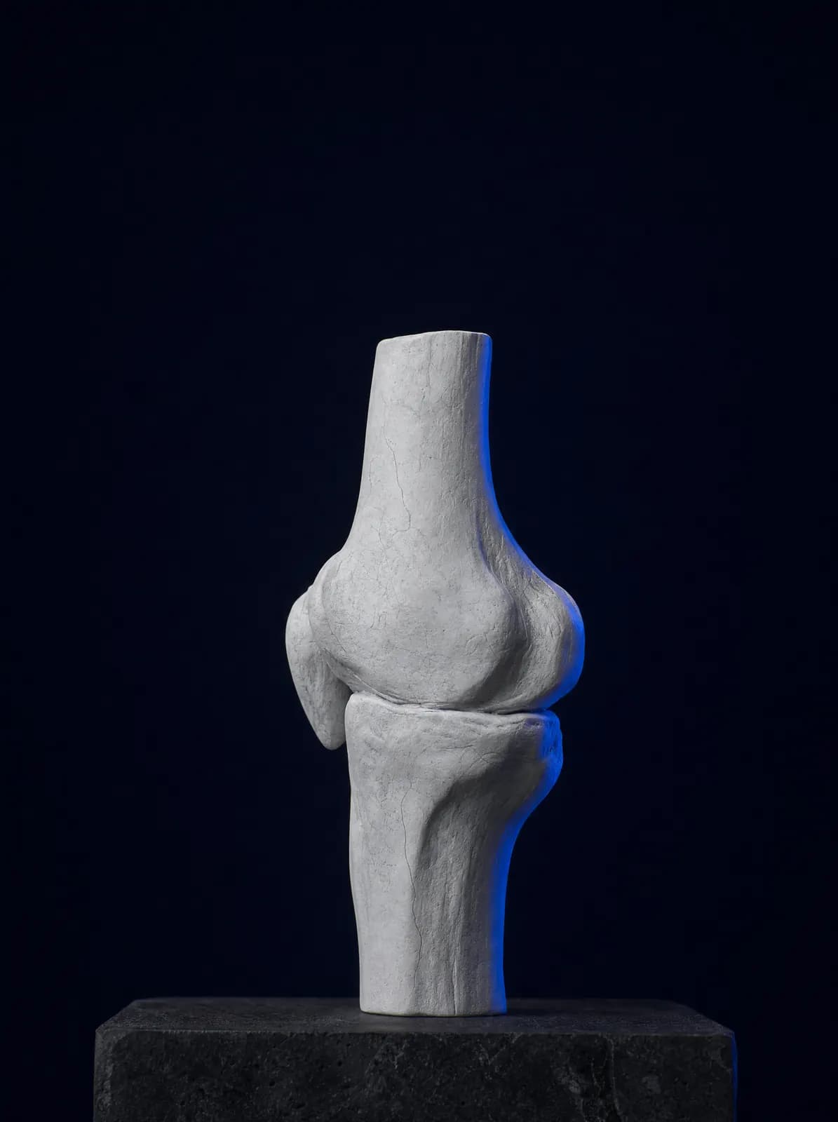
The knee joins the thighbone (femur), shinbone (tibia) and kneecap (patella), with joint surfaces covered in cartilage. Cartilage allows the bones to glide without rubbing together. Osteoarthritis is gradual thinning and surface damage of this cartilage; bony spurs (osteophytes) form at the edges, the joint lining becomes irritated and fluid may accumulate.
Although the Turkish term commonly used means 'calcification', calcium does not literally build up in the joint; the name comes from the bony spurs visible on X-ray. It is most common after age 50. Excess weight, previous meniscal or ligament injuries, intra-articular fractures, knock knees or bow legs, and a family history increase risk.
What Are the Symptoms?
Symptoms usually develop slowly and increase over time. Patients most commonly describe:
- Knee pain that increases when going up or down stairs, squatting and standing up
- Stiffness lasting a few minutes in the morning or after prolonged sitting
- Knee swelling and a grinding or crackling sensation with movement
- Reduced walking distance; night pain at an advanced stage
- Inability to fully straighten or bend the knee; increasing leg angulation
How Is It Diagnosed?
Diagnosis uses examination and standing knee X-rays, assessing joint-space narrowing, bony spurs and leg alignment. MRI is not needed for everyone; it is requested for suspected meniscal tears, assessment of cartilage damage or another possible cause. Blood tests do not diagnose osteoarthritis but may help distinguish rheumatic disease when needed.
Examination assesses pain location (inner, outer or front), movement, ligament stability and hip-to-ankle alignment together. Planning depends on limitations in daily life as well as the X-ray stage.
Stages and Treatment Decisions
Osteoarthritis is classified into four radiographic stages (Kellgren–Lawrence). In early stages (1–2), the joint space is largely preserved; the aim is to slow progression and control pain. In the moderate stage (3), pain begins to affect daily life, and the limits of non-surgical methods are discussed. In the advanced stage (4), bone contacts bone; the most frequently discussed surgical option at this point is knee replacement.
Decisions depend on age, weight, expectations and other conditions as well as the X-ray stage. Two patients with identical X-rays may receive different plans; the reasons are explained clearly during consultation.
Non-Surgical Treatments
Non-surgical treatment rests on three measures relevant at every stage: weight control, muscle-strengthening exercises, particularly for the quadriceps, and activity modification. Painkillers, anti-inflammatory medicines, physiotherapy and, when needed, joint injections are added accordingly.
Intra-articular procedures include hyaluronic acid, sometimes called a rooster-comb injection, PRP, cortisone and regenerative methods such as stem cells, exosomes, polynucleotides and chitosan. Their relevance varies by disease stage and patient; evidence levels, number of sessions and expected duration of effect are compared on the regenerative and non-surgical treatments page.
Treatment of Knee Osteoarthritis in Older People
In older adults, the main aim is usually to preserve walking distance and independence. Exercises are adapted to fall risk and injections selected according to coexisting conditions; surgery is based on general health and expectations rather than age. For general information, see the orthopaedics in older adults page.
In patients over 65, osteoarthritis is not assessed in isolation. High blood pressure, diabetes, heart disease and anticoagulant use affect both medicine selection and injection planning. Doses and treatment duration are limited because painkillers can affect the stomach and kidneys; non-drug measures such as exercise, reduced loading, suitable shoes and a walking stick when needed therefore become even more important. A correctly adjusted stick held in the hand opposite the painful knee can substantially reduce loading on the knee.
Fall risk is another important issue in older age. Knee pain alters walking, muscles weaken and balance deteriorates; a fall may then cause a much more serious injury such as a hip fracture. Balance exercises, home modifications such as securing rug edges, bathroom grab rails and lighting, and vitamin D and bone-health assessment are therefore added to the programme. Patients with osteoporosis receive a separate assessment of this.
Age alone does not prevent surgery. Joint replacement may be recommended for an 80-year-old in suitable general health, while non-surgical care may be preferred for a younger person with severe coexisting disease. The deciding factors are not age but the extent to which pain limits daily life, the patient's expectations and their fitness for surgery.
What Is Patellofemoral Osteoarthritis?
Osteoarthritis does not always start in the inner or outer compartment of the knee. In some patients, wear mainly affects the surface between the kneecap and thighbone (the patellofemoral joint). Pain is at the front of the knee and becomes worse when descending stairs, walking downhill, standing after prolonged sitting or squatting. Patients often describe pain "at the front of my knee, behind the kneecap"; crackling and a grinding sensation may accompany movement.
Kneecap osteoarthritis often involves poor tracking: a tendency for the kneecap to move outwards, weakness in the inner quadriceps, insufficient hip muscle control or a previous kneecap dislocation. Assessment therefore includes hip and foot alignment as well as the knee; special X-ray views showing the kneecap's track may be requested.
Exercise is the mainstay of treatment: strengthening the quadriceps and outer hip muscles, improving flexibility and modifying movements that increase loading, such as deep squatting, prolonged stair use and kneeling. Taping or kneecap-supporting braces may provide temporary relief in selected patients. If symptoms persist despite non-surgical treatment and wear is confined to this area, surgical options are assessed separately; decisions combine examination and imaging findings.
What Should the Exercise Programme Include?
Exercise is widely discussed but often not maintained in osteoarthritis. Its aim is not to tire the knee but to strengthen the muscles supporting it. Strong quadriceps share joint loading, while hip muscles prevent the knee collapsing inwards during walking. These two muscle groups form the programme's foundation.
Initially, choose low-stress movements that do not increase pain: straight-leg raises lying on your back, slowly straightening the knee while seated, shallow wall squats and side-lying leg raises. Perform movements slowly; consistency matters more than repetitions. A programme of 10–15 minutes on most days, continued for months, is more helpful than an intensive programme abandoned after two weeks.
Low-impact activities such as walking, swimming and stationary cycling complement the programme. On a stationary bicycle, adjust the saddle so the knee remains slightly bent at the bottom of the pedal stroke; keep resistance low and the pace comfortable. Treadmill inclines may increase pain at the front of the knee. Mild effort during exercise is normal, but pain lasting into the next day or causing swelling means the programme is too demanding and loading should be reduced.
When swelling is present, it is controlled before strengthening begins. Suitable movements depend on knee range, disease stage and previous surgery; the programme is personalised during examination and coordinated with physiotherapy when necessary.
When Is Surgery Needed?
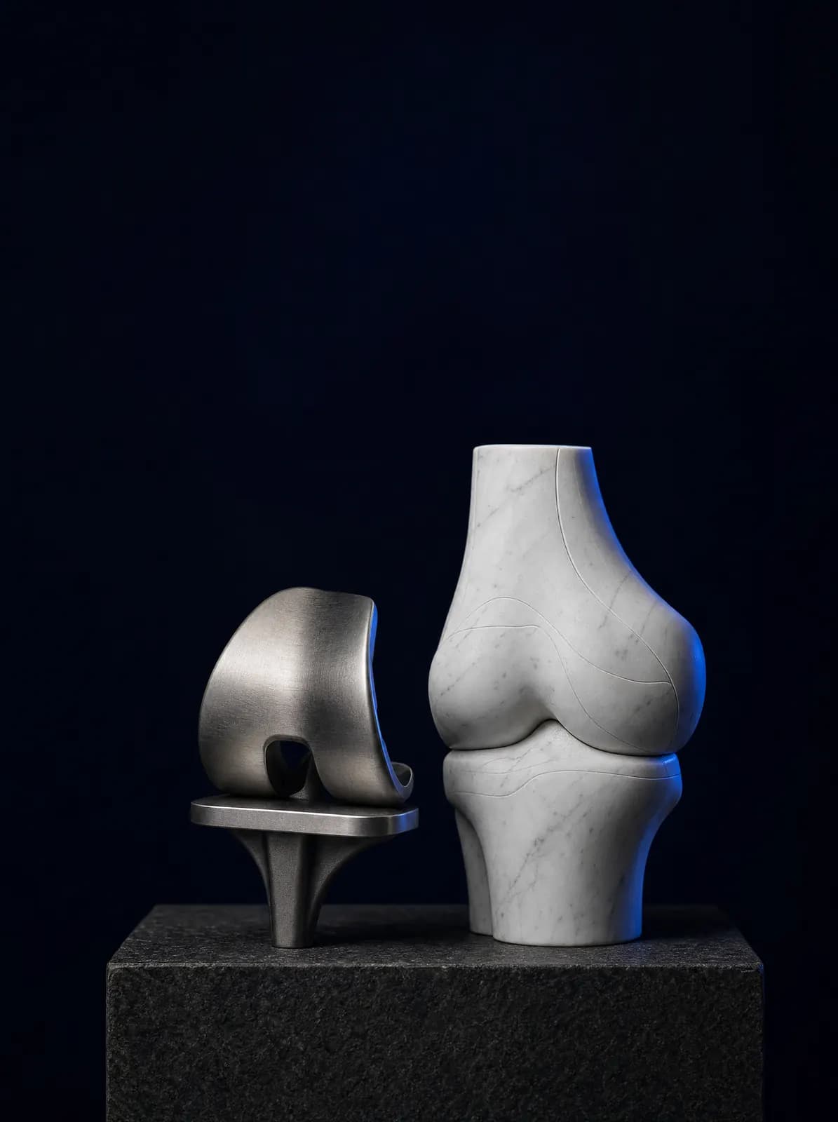
Surgery is discussed when pain disrupts sleep despite non-surgical care, walking distance is markedly reduced and X-rays show advanced narrowing. Options vary: realignment (osteotomy) for younger patients with marked deformity, partial replacement when only one compartment is worn, and total knee replacement for widespread wear.
The operation, hospital stay, postoperative walking and follow-up are explained in detail on the knee replacement surgery and recovery page.
What Should You Consider in Daily Life?
Regular joint-friendly activity such as walking and swimming protects the knee; prolonged squatting, cross-legged sitting and stairs increase loading. Each 1 kg of body weight produces approximately 3–4 times that load at the knee during walking; even modest weight loss may reduce pain. Suitable shoes, a walking stick if needed and consistent home exercise are part of treatment.
Frequently Asked Questions
Worn cartilage does not spontaneously return to its original state. Treatment aims to slow progression, control pain and preserve movement; effects of regenerative methods on cartilage remain under investigation and are discussed with their evidence level at consultation.
An orthopaedics and traumatology specialist provides diagnosis and treatment planning, working with physiotherapy for exercises.
Usually yes in early and moderate stages: weight control, exercise, medicines and joint injections may manage pain. Their effect is limited in advanced disease; options are assessed at consultation.
Corticosteroid injections are limited in number and spaced apart; hyaluronic acid and PRP schedules vary with product and patient. Which injection and how often are decided at consultation.
No. Regular low-load activity, such as level-ground walking and swimming, is recommended. Squatting, stairs and prolonged kneeling are more demanding.
You can book by phone or WhatsApp; bringing previous X-rays or MRI images helps assessment.
References

Appointments and Contact
Call or contact us on WhatsApp to arrange an appointment. Briefly describe your symptoms; we try to schedule your examination and any necessary imaging on the same day.
- Opening hours
- 08:30–18:00Monday – Saturday
The clinic is not a 24/7 emergency department. For a fracture, dislocation, open wound, or inability to bear weight with fever, call 112 or attend the nearest hospital emergency department.