Meniscal Tear: Non-Surgical Treatment and Arthroscopic Repair
The meniscus is crescent-shaped cartilage that acts as a knee shock absorber. Not every tear needs surgery; this page explains when non-surgical monitoring and when arthroscopic repair are considered.
Also known as: Meniscal lesion, Meniscal disorder, Meniscal rupture
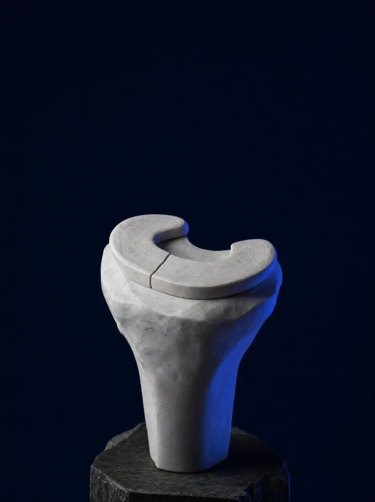
Your Treatment Pathway
Examination
A preliminary diagnosis is made from the history, joint-line tenderness and rotational tests.
Imaging
X-rays exclude other causes; MRI defines tear type when necessary.
Non-Surgical Programme
Activity modification, swelling control and strengthening, usually tried for 6–8 weeks.
Arthroscopy If Necessary
Repair for suitable tears; removal of as little tissue as possible when healing is unlikely.
Movement and Follow-up
Return to daily life and sport with gradual loading, strengthening and balance exercises.
What Is a Meniscal Tear?
There are two menisci, medial and lateral, between the thigh bone and shin bone in the knee. These flexible, crescent-shaped cartilage structures distribute load over a wide area, absorb impact and help the joint move smoothly. A tear in this tissue is called a meniscal tear.
Tears broadly fall into two groups. Traumatic tears occur in young, active people when the knee twists suddenly with the foot planted; football, small-sided football, skiing and rising from a squat are common causes. Degenerative tears occur after middle age as meniscal tissue wears over time, sometimes after only minor strain, and frequently accompany knee osteoarthritis. This distinction matters because the treatment pathways differ.
The outer meniscal edge has a blood supply and a relatively better chance of healing; supply decreases towards the inner edge, where spontaneous healing is less likely. Tear location, shape (longitudinal, radial, bucket-handle, flap or root) and size directly influence treatment.
What Are the Symptoms?
Symptoms vary with tear type. A traumatic tear often has a clearly remembered onset, while a degenerative tear develops gradually.
- Pain felt along the inner or outer joint line of the knee
- A catching sensation that increases with squatting, turning and walking downstairs
- Catching, occasional locking and inability to fully straighten the knee
- Swelling developing within hours of the injury, followed by recurrent joint fluid accumulation
- Lack of confidence in the knee and sudden giving way, which may resemble ligament injury
How Is It Diagnosed?
Diagnosis rests on the history and examination. Joint-line tenderness, tests rotating the knee at particular angles and movement restriction are assessed. X-rays do not show the meniscus but are obtained initially to assess osteoarthritis and joint space and exclude other causes such as fracture.
MRI (magnetic resonance imaging) shows the location and shape of a meniscal tear. However, seeing a tear on MRI does not by itself mean surgery is needed: meniscal changes are also common in symptom-free knees in people beyond middle age. The decision therefore considers examination findings and the effect of symptoms on daily life together, rather than the MRI report alone.
Can a Meniscal Tear Resolve Without Surgery?
Many meniscal tears, particularly degenerative ones, can be managed without surgery. The aim is to reduce pain, control swelling and restore safe knee use. During the first weeks, aggravating movements such as deep squatting, pivoting and extensive stair use are limited, with ice and anti-inflammatory medication when needed.
The key step is an exercise programme: strengthening the front thigh muscle (quadriceps) and muscles around the hip transfers load from the meniscus to the muscles. The programme continues over weeks with a physiotherapist or through a prescribed home programme. If swelling and pain persist, intra-articular procedures may be considered for selected patients; suitability, evidence levels and session plans are compared on the regenerative and non-surgical treatments page.
Non-surgical care is generally assessed over a six–eight-week trial. If symptoms improve substantially, the programme continues; persistent locking or pain that still restricts daily life prompts renewed discussion of surgery.
When Is Arthroscopic Surgery Needed?
Surgical decisions depend on tear type and the patient's situation. Main indications include true locking preventing full extension, especially a bucket-handle tear, traumatic tears in young patients, meniscal root tears and persistent symptoms despite non-surgical rehabilitation.
Surgery is performed arthroscopically: a camera and fine instruments enter the knee through two–three openings a few millimetres wide. There are two main approaches. Repair (stitching) is preferred when the tear lies in a region with a blood supply and the tissue is healthy; it preserves the meniscus, but recovery is longer and weight-bearing and bending are restricted in the first weeks. Partial meniscectomy removes a torn portion that cannot heal, taking away as little tissue as possible; recovery is faster, but some meniscal tissue is lost.
Preserving as much meniscus as possible is essential because reduced meniscal tissue increases loading on joint surfaces over the long term. Repair is therefore considered first whenever suitable. The full extent of surgery sometimes becomes clear only after entering the knee; the possibilities are discussed beforehand.
Meniscal Tears in Children and Adolescents
Meniscal tissue has a better blood supply in children and adolescents, so repair generally has a higher chance of healing than in adults. The approach in this age group is strongly preservation-focused: wherever possible, the meniscus is sutured and retained, with tissue removal a last resort. Losing meniscal tissue at a young age may contribute to faster wear of the joint surfaces later in life.
Another consideration in this age group is a meniscus that is disc-shaped from birth rather than crescent-shaped (discoid meniscus). This can cause catching, audible movement and locking without a significant injury. Repeated knee locking, a pronounced sound during movement or an unexplained limp in your child should be assessed by examination. For other orthopaedic conditions specific to children, see paediatric orthopaedics.
How Does Recovery Progress After Surgery?
Recovery depends on the procedure. After partial meniscectomy, most patients walk with aids during the first days and return to daily activities within a few weeks as swelling settles. After meniscal repair, the sutured tissue must heal: weight-bearing and bending are kept within limits set by your doctor during the first weeks, and a brace may be used temporarily.
In both cases, rehabilitation follows the same sequence: controlling swelling, restoring full extension, gradual bending, strengthening and finally balance and coordination. Return to sport is gradual once strength and control tests are satisfactory; achieved function, rather than the calendar, determines readiness. Returning too early or without control risks re-tearing the repaired meniscus.
What Should You Consider in Daily Life?
Movements that most strain a knee with meniscal problems include twisting with the foot planted, deep squatting, sitting cross-legged and prolonged kneeling. Walking on level ground, swimming and stationary cycling are recommended instead. Weight control directly reduces knee load.
Knees with reduced meniscal tissue may have an increased long-term risk of cartilage wear; exercises that maintain muscle strength should therefore continue throughout life. For symptoms and treatment stages, see the knee osteoarthritis page. You can book by telephone or WhatsApp at AC CLINIC in Altınordu, Ordu, for examination of meniscal symptoms and planning of the necessary imaging.
Frequently Asked Questions
Some tears, particularly degenerative tears after middle age, can be managed without surgery through exercise and activity modification. Surgery is more prominent for true locking, bucket-handle tears or root tears. Examination and imaging establish the distinction.
Surgery is arthroscopic, through two–three openings a few millimetres wide in the knee. Their scars gradually fade. Repair sometimes requires a small additional incision to place stitches.
Desk work is usually possible within a few days to two weeks after partial meniscectomy; standing or heavy work takes longer. Recovery after meniscal repair is longer. Return to sport is gradual and guided by strength and control tests rather than a calendar.
An MRI finding alone is not a reason for surgery. Meniscal changes are common after middle age even in people without symptoms. Decisions depend on locking, how much pain limits daily life and examination findings.
Only the outer edge of the meniscus has a good blood supply, so small tears there may heal. Tears at the inner edge are not expected to heal spontaneously because blood supply is poor; treatment then aims to control symptoms and protect the knee.
You can book an examination by phone or WhatsApp at AC CLINIC in Altınordu. Bringing any knee X-rays and MRI images on CD or reports helps assessment.
References
Appointments and Contact
Call or contact us on WhatsApp to arrange an appointment. Briefly describe your symptoms; we try to schedule your examination and any necessary imaging on the same day.
- Opening hours
- 08:30–18:00Monday – Saturday
The clinic is not a 24/7 emergency department. For a fracture, dislocation, open wound, or inability to bear weight with fever, call 112 or attend the nearest hospital emergency department.