Osteoporosis: Diagnosis and Treatment
Osteoporosis weakens bone density and internal structure, making fractures more likely. It progresses silently; for many patients, the first sign is a fracture after a minor fall. This page explains diagnosis and how treatment is planned to reduce fracture risk.
Also known as: Osteoporosis, Osteoporosis, Loss of bone density
Your Treatment Pathway
Assessment
Fracture history, height loss, medicines, fall risk and dietary habits are reviewed.
Measurements and Tests
Bone density measurement (DEXA), X-rays if needed and blood tests investigating the underlying cause.
Plan
Calcium and vitamin D intake, exercise and medication if fracture risk requires it are planned together.
Review
Medication adherence, side effects, height and posture are monitored; scans are repeated at doctor-determined intervals.
Movement
Aim: prevent falls and further fractures while preserving independence in daily life.
What Is Osteoporosis?
Bone is living tissue that renews throughout life, not an inert structure. Old bone is continually broken down and replaced. Formation exceeds breakdown in youth, with peak bone mass reached at approximately 25–30 years. The balance then gradually shifts towards breakdown. Osteoporosis occurs when this loss progresses enough to make bones prone to fracture.
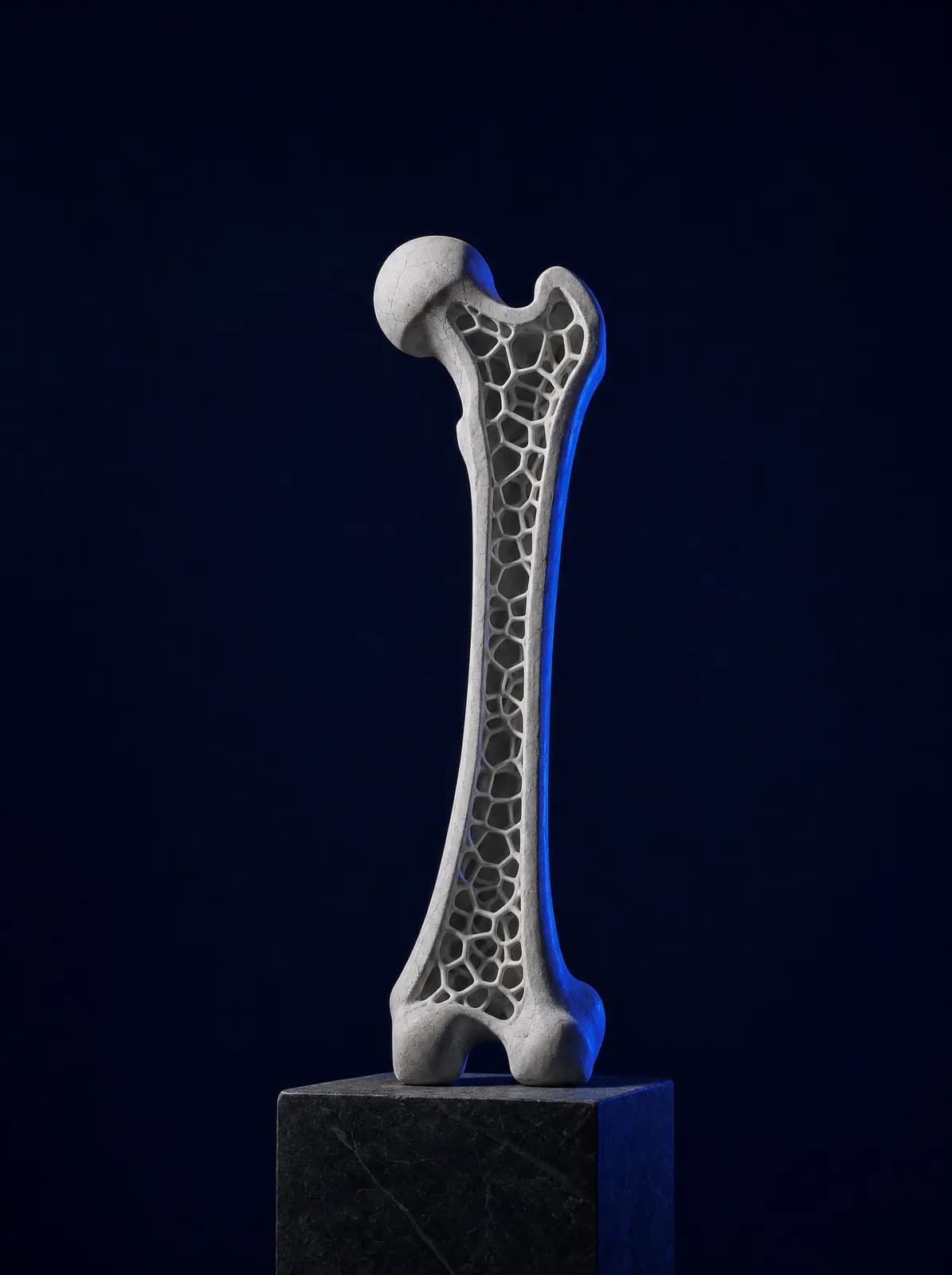
The problem is more than "less bone". The honeycomb-like internal network thins and loses connections, making bone both less dense and less resistant to impact. A fall that would not harm a healthy bone may therefore break an osteoporotic one. This is called a fragility fracture: a fracture caused by low-energy trauma, such as a fall from standing height.
The intermediate stage of bone loss that has not reached the osteoporosis threshold is called osteopenia (low bone mass). Osteopenia alone is not considered a disease, but it informs treatment decisions when other fracture risk factors are present. Bone loss accelerates in the first years after menopause, but osteoporosis also affects men and is often overlooked in them.
What Are the Symptoms?
The difficult aspect of osteoporosis is that it may cause no symptoms for a long time, hence the name "silent disease". Bone loss itself does not cause pain. Widespread body pain is usually due to another condition, while someone with no pain may have substantial bone loss. The condition is often discovered after a fracture or during routine screening.
- A fracture after a minor fall, sneeze or heavy lifting, most commonly affecting the wrist, hip or spine
- Loss of height over the years by more than a few centimetres
- Forward bending of the back and a markedly hunched posture
- Sudden severe back or lower back pain that increases with movement may indicate a vertebral compression fracture
- Ribs moving closer to the pelvis, the abdomen protruding forwards and a feeling of breathlessness
Who Is More Commonly Affected?
Some risks cannot be changed, while others relate to daily habits. Both groups are reviewed because treatment decisions depend on overall fracture risk, not the measurement alone.
- Increasing age; menopause in women, particularly early menopause before age 45
- A parental history of hip fracture, a thin build and low body weight
- A previous fracture following minor trauma (one of the strongest warning signs)
- Long-term cortisone (corticosteroid) use and certain medicines
- Smoking, excessive alcohol, inactivity and being bedbound
- Vitamin D deficiency, a diet low in calcium and limited sun exposure
- Thyroid and parathyroid disease, rheumatoid arthritis, malabsorption such as coeliac disease, and chronic kidney or liver disease
How Is It Diagnosed, and What Is Bone Density Measurement?
Assessment starts with history and examination: previous fractures, height loss, medicines, falls and diet are discussed. The main diagnostic tool is bone density measurement (DEXA / DXA). This painless scan uses low-dose X-rays, takes approximately 10–15 minutes and usually measures the lumbar spine and hip.
The result is reported as a T-score. The generally accepted classification is: −1 or above is normal, between −1 and −2,5 is low bone mass (osteopenia), and −2,5 or below is osteoporosis. In someone who has sustained a fragility fracture, osteoporosis may be diagnosed even when the measurement is borderline. Tools estimating fracture risk using age, sex, fracture history and other factors also inform the decision.
X-rays are not sensitive enough to show early bone loss, but they can identify vertebral compression fractures. Blood and urine tests, including calcium, phosphate, vitamin D, thyroid and parathyroid hormones, and kidney and liver function, investigate other diseases causing bone loss. This distinction matters because an underlying cause becomes the first treatment target.
What Are the Treatment Options?
The goal is preventing fractures, not merely changing a bone density number. The plan has three parts: diet and supplements, exercise and fall prevention, and medication where needed.
Basic Measures: Calcium, Vitamin D and Exercise
Calcium should come primarily from food: milk and dairy products, green leafy vegetables, pulses, sesame and grape molasses. Adequate protein also matters for maintaining muscle mass. Vitamin D is measured through a blood test, and deficiency is corrected at a dose determined by the doctor; taking high-dose supplements indiscriminately is not appropriate.
Exercise benefits bone because loading strengthens it. Walking, light resistance training and balance exercises are recommended together; swimming benefits the heart and lungs but is not sufficient alone because it does not load bone. The programme is adapted to coexisting conditions and fall risk.
Who Needs Medication?
Decisions about medication consider bone density measurements, previous fractures, age and overall fracture risk together. The most commonly used group is bisphosphonates, available as weekly or monthly tablets or an annual intravenous treatment. Other options include an injection under the skin every six months that suppresses bone breakdown, and medicines that stimulate bone formation in selected patients at high risk of fracture. Menopause-related treatments are assessed jointly with a gynaecologist.
All these medicines have instructions for use and possible side effects: oral bisphosphonates are taken on an empty stomach with plenty of water, remaining upright for a period afterwards; because of rare jawbone problems, doctors must be informed before planned tooth extractions or implant procedures. A treatment break (a "drug holiday") is considered only with bisphosphonate treatment and on a doctor's decision; it does not apply to every medicine. Missing a dose of the injection given every six months or stopping it without medical advice can lead to rapid bone loss and spinal fractures, so any decision to stop must be made with the doctor, usually with a switch to another medicine. Medicines should therefore not be started without a prescription, changed on a neighbour's advice or continued without regular review.
After a Fracture: Preventing Another Fracture
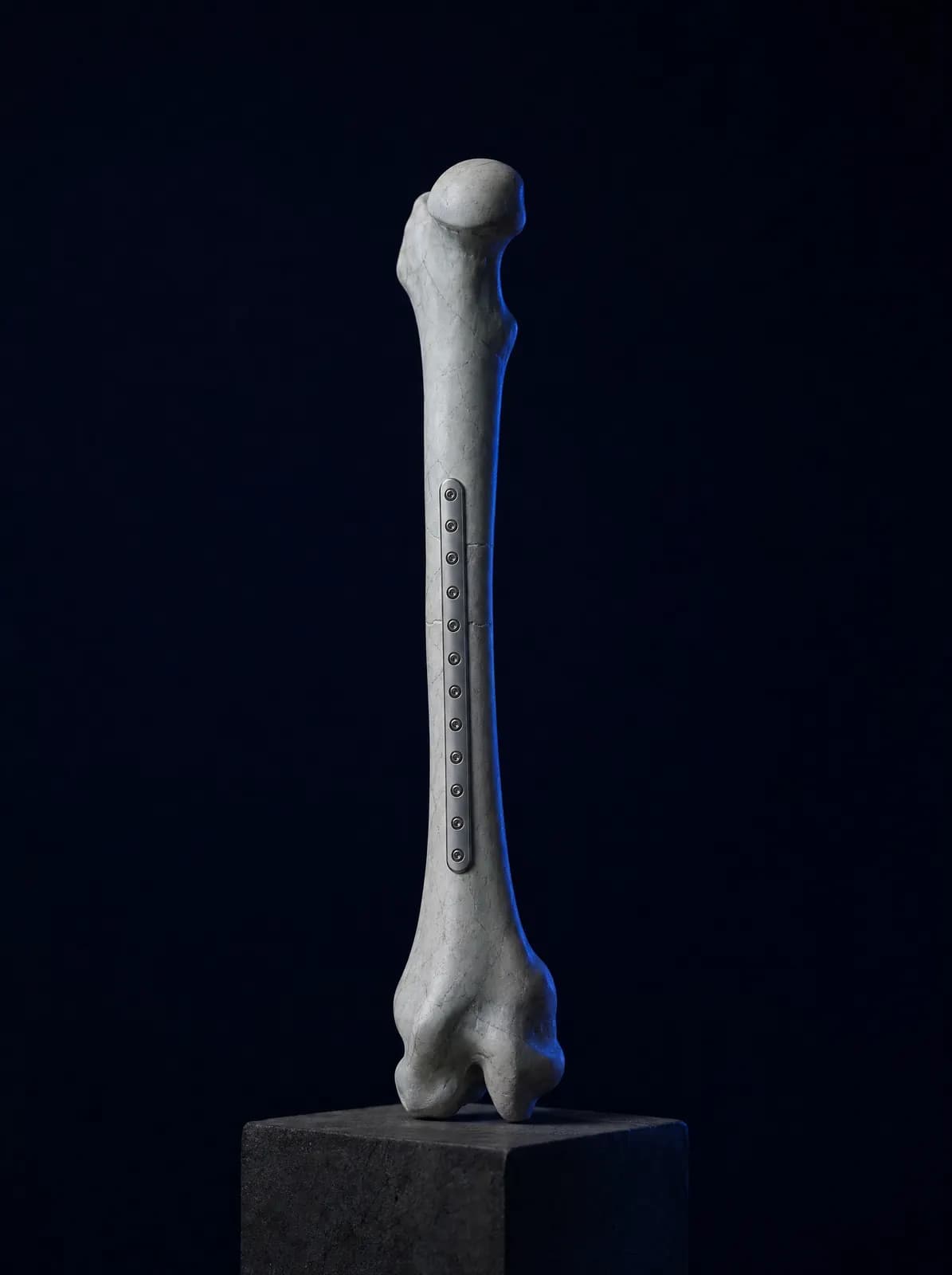
Fracture treatment itself, whether casting, fixation or replacement, depends on its location and type and is described on the relevant pages. This section addresses the step often missed after fracture treatment: investigating and treating why the bone broke so easily.
A fragility fracture is a strong indication of osteoporosis. Someone who has had one has a markedly higher risk of another fracture than a person of the same age without a previous fracture, particularly during the first year. A fracture is therefore now viewed not as an isolated event but as a point at which bone health needs reassessment.
In practice, this means arranging a bone density scan while the fracture heals or soon afterwards, checking vitamin D and calcium levels and screening for other conditions that can cause bone loss, starting medication in suitable patients and reviewing the causes of falls. A history of fracture may alter the treatment decision even if the scan result is borderline. Because immobility after a fracture accelerates bone loss, early movement and rehabilitation within the limits permitted by the doctor are also part of this plan.
This assessment should not be delayed even if the fracture was treated elsewhere; bring X-rays and discharge summaries from that period to help planning.
What Should You Consider in Daily Life?
Daily habits are part of osteoporosis treatment. The points below directly concern bone and spinal health. Since most fractures follow a simple fall at home, home safety and fall prevention are equally important; a detailed list is in the walking difficulties and falls in older adults section.
- Obtain calcium primarily from food: milk and dairy products, leafy greens, pulses, sesame and molasses
- Have vitamin D measured and take the dose prescribed if deficient; do not take high doses indiscriminately
- Eat enough protein at every meal; preserving muscle mass goes hand in hand with preserving bone
- Stop smoking and limit alcohol; both accelerate bone loss
- Do not lift heavy loads by bending at the waist; bend the knees, keep the load close to your body and avoid twisting your trunk
- Avoid sit-up-type exercises that bend the spine forwards and sudden trunk rotation; ask your doctor about your programme, particularly after a vertebral fracture
- Make regular weight-bearing walking and strengthening and balance exercises two to three days a week a habit
Frequently Asked Questions
An orthopaedics and traumatology specialist assesses fractures, fracture risk and the skeletal system. If a hormonal or metabolic cause is suspected, endocrinology is involved; inflammatory rheumatic disease requires rheumatology, and exercise and rehabilitation programmes are coordinated with physiotherapy. What matters is not which specialty you first attend, but maintaining treatment consistently.
The scan is painless, takes approximately 10–15 minutes and uses a low radiation dose. It is commonly requested for postmenopausal women, older men, people with a fracture after minor trauma, prolonged corticosteroid users and those with a family history of hip fracture. Individual risk assessment determines who needs it and how often.
Calcium and vitamin D provide the foundation of treatment, but are insufficient alone to prevent fractures in established osteoporosis. Medication is added for patients at high fracture risk. Supplements should not be used indiscriminately: vitamin D dosing depends on blood levels, and calcium dosing accounts for dietary intake.
The duration depends on the medicine, fracture history and response to treatment; it is generally measured in years and reviewed at intervals. A treatment break (a "drug holiday") is considered only for people taking bisphosphonates and on a doctor's decision. The injection given every six months is different: delaying a dose or stopping treatment without medical advice can accelerate bone loss and lead to spinal fractures within a short time. The decision to stop is therefore made with the doctor, usually with another medicine planned in its place. Do not stop any osteoporosis medicine yourself without a review appointment.
Walking is a good start but is not sufficient alone. Weight-bearing activity to strengthen bone, light resistance work to preserve muscle and balance exercises to reduce falls are recommended together. Movements that bend the spine forwards or strain the trunk should be avoided, particularly after a vertebral fracture; the programme is tailored individually.
Yes. It occurs later in men than women, but outcomes after fracture may be more severe. Long-term corticosteroids, smoking, alcohol, low testosterone and chronic disease are important causes in men; testing should not be delayed when these risks are present.
You can book by phone or WhatsApp; AC CLINIC is in Altınordu, Ordu. Bringing previous bone density results, recent blood tests and a medication list helps assessment.
References

Appointments and Contact
Call or contact us on WhatsApp to arrange an appointment. Briefly describe your symptoms; we try to schedule your examination and any necessary imaging on the same day.
- Opening hours
- 08:30–18:00Monday – Saturday
The clinic is not a 24/7 emergency department. For a fracture, dislocation, open wound, or inability to bear weight with fever, call 112 or attend the nearest hospital emergency department.