Developmental Dysplasia of the Hip (DDH) in Babies: Screening, Ultrasound and Treatment
Developmental dysplasia of the hip means the baby's hip is underdeveloped or dislocated. When detected early through examination and ultrasound, treatment often uses a simple harness; later diagnosis may require casting or surgery.
Also known as: Developmental dysplasia of the hip (DDH), Congenital hip dislocation, Hip dysplasia, DDH (developmental dysplasia of the hip)
Your Treatment Pathway
Examination
The baby's hip examination, risk factors and family history are assessed together.
Imaging
Hip ultrasound or X-rays, depending on age, are used to assess socket development.
Plan
A harness, cast or surgical pathway is chosen according to age and findings, addressing the family's questions.
Administration
The device or cast is fitted, care instructions provided and positioning confirmed with imaging.
Growth Monitoring
Aim: regular monitoring of socket development and walking throughout growth.
What Is Developmental Dysplasia of the Hip?
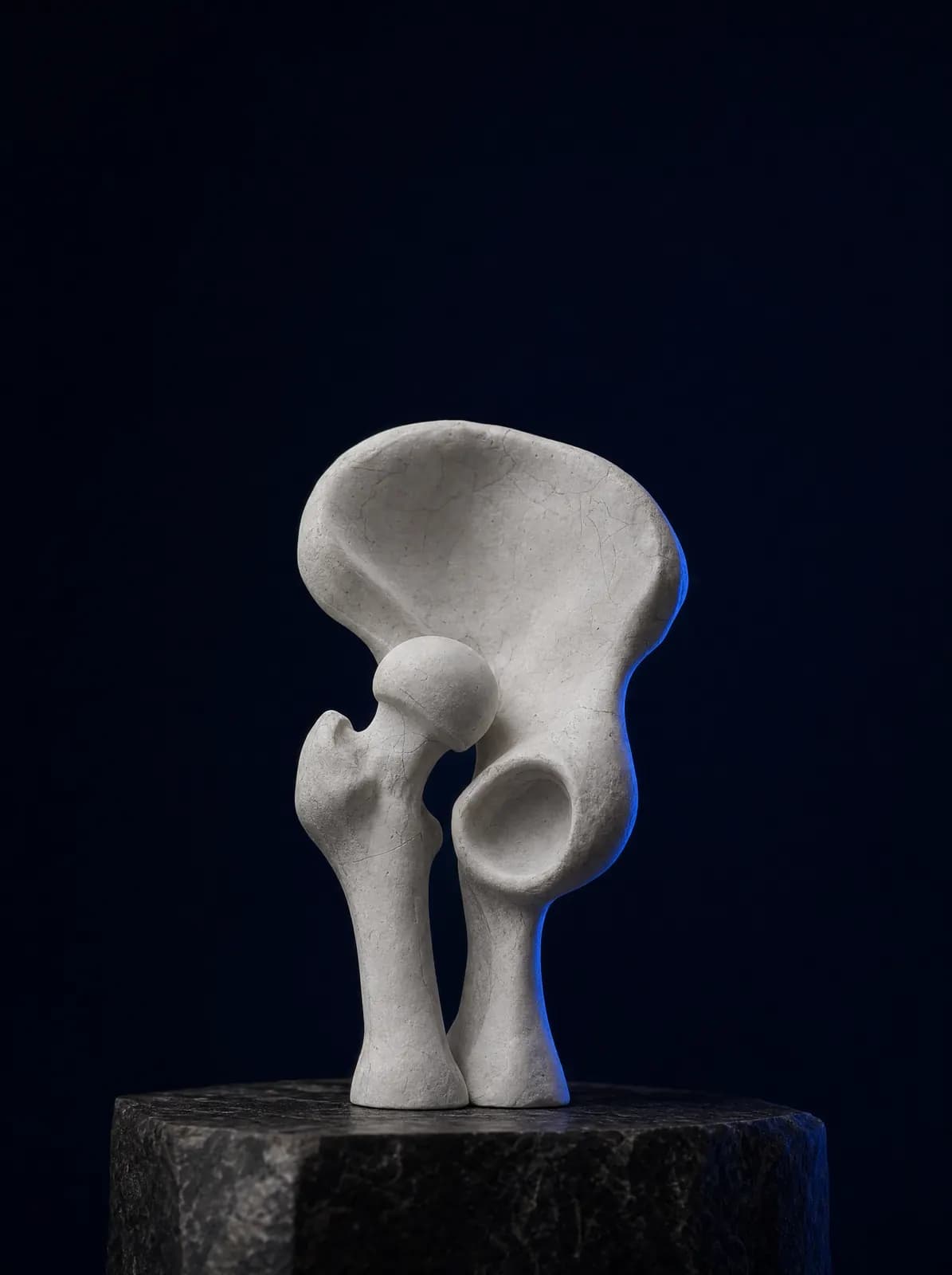
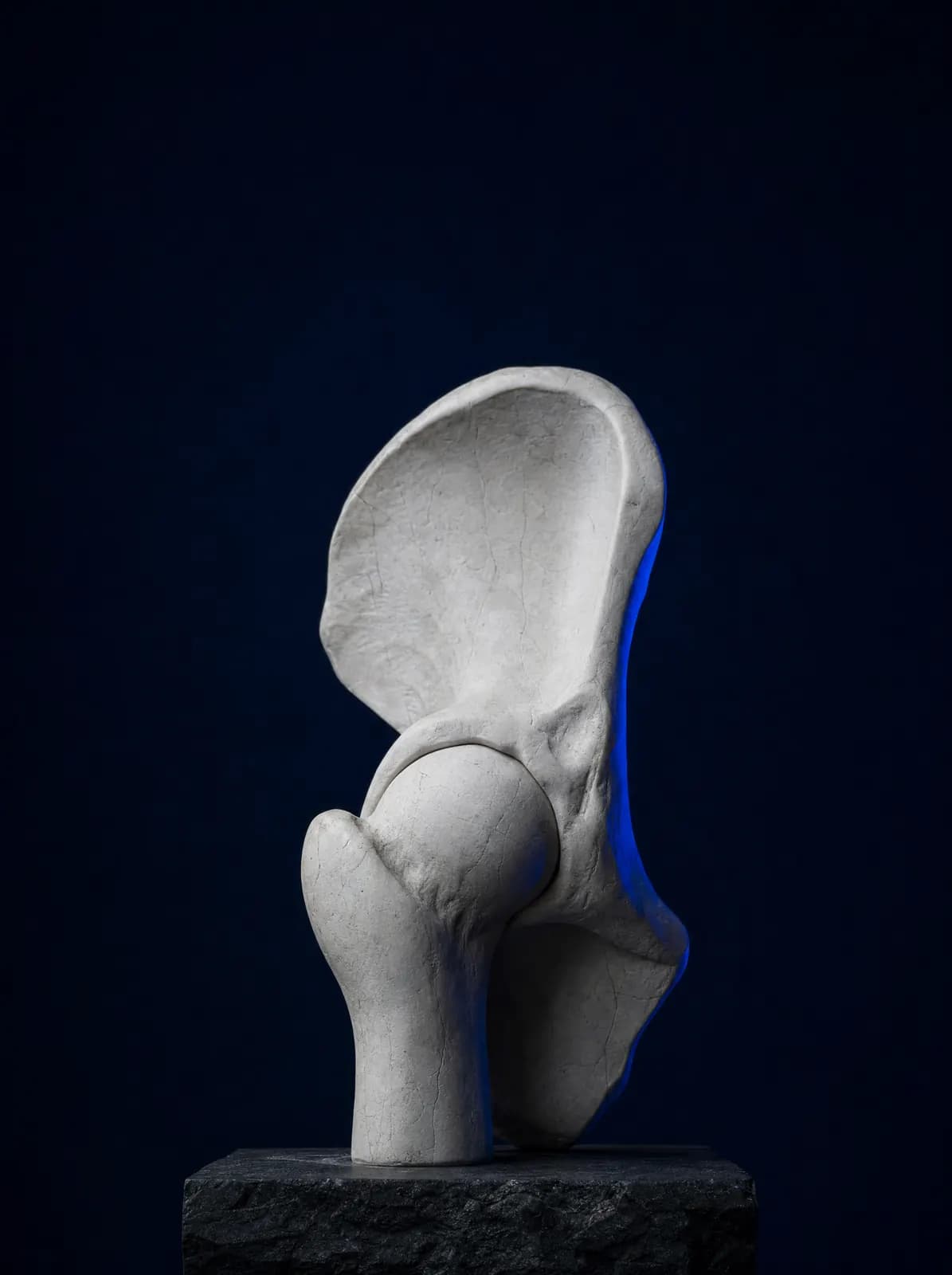
The hip joint consists of the ball at the upper end of the femur (femoral head) and the socket in the pelvis (acetabulum). In babies, the socket is still soft and largely cartilaginous; the ball must stimulate it correctly for it to deepen. Developmental dysplasia of the hip (DDH) disrupts this relationship: the socket remains shallow and the ball slips partly or completely out.
It is commonly called "congenital hip dislocation", but doctors use "developmental" because it is not always present at birth and may appear or worsen in the first months. One newborn examination is therefore insufficient; repeated early checks matter.
DDH is a spectrum rather than a single condition: a shallow socket (dysplasia), partial displacement of the ball (subluxation) or complete dislocation. Position within this spectrum affects both treatment and follow-up duration.
How Is It Recognised in Babies? What Are the Signs?
Most babies with developmental dysplasia of the hip (DDH) have no signs that families can recognise. They do not cry or become unsettled and move their legs normally. Its painless nature makes early screening essential. Nevertheless, the following signs warrant prompt orthopaedic assessment:
- One leg is difficult to open outwards during nappy changing
- Marked asymmetry between the skin folds of the legs and hips
- One knee sitting lower than the other when the baby lies on their back with knees bent, suggesting unequal leg lengths
- A snapping or clicking sensation heard or felt when moving the leg
- Waddling, limping or one-sided toe walking in a child beginning to walk
- A pronounced lower-back curve and legs held apart in bilateral cases
Which Babies Are at Greater Risk?
DDH can affect any baby, but certain factors substantially increase frequency. Even without symptoms, hip ultrasound is planned if any of the following applies:
- Breech presentation at birth
- A family history of hip dislocation (mother, father, sibling)
- Being female and firstborn
- Low amniotic fluid (oligohydramnios) and multiple pregnancy
- Associated congenital foot deformities or a twisted neck (torticollis)
- Tight swaddling that keeps the legs together and straight
How Is the Diagnosis Made, and When Is Hip Ultrasound Performed?
Examination is the first diagnostic step. In newborns, specific manoeuvres (Ortolani and Barlow tests) assess whether the hip dislocates; in later months, hip abduction and leg length difference are evaluated.
Age determines the imaging method. In the first months, much of the infant hip is cartilage and is not clearly visible on X-ray, so hip ultrasound is used. Ultrasound is generally useful after the first weeks of life, commonly from weeks 4–6 until month 6. After approximately 4–6 months, as ossification progresses, assessment uses an anteroposterior hip X-ray.
The timing and number of repeat ultrasound scans depend on the baby's examination and risk factors. Following assessment at AC CLINIC in Ordu, suitable hip imaging is arranged; bringing any previous ultrasound or X-ray images is useful for comparison.
What Are the Treatment Options?
Treatment has one aim: place the femoral head in the socket and keep it there so the socket can develop normally. The method depends on the baby's age, whether the hip is dislocated or merely shallow, and previous treatment. Generally, the earlier the diagnosis, the simpler the treatment.
0–6 Months: Harness and Brace Treatment
The Pavlik harness and similar devices used in the first months of life hold the legs flexed at the hips and apart. This position allows the ball to sit in the socket. The device is worn throughout the day for a specified period, and ultrasound at follow-up checks positioning. Parents receive detailed advice on skin care, nappy changing and dressing; they should not alter the harness settings themselves.
6–18 Months: Closed Reduction and Casting
If a harness does not achieve reduction or diagnosis is late, the hip may be reduced under anaesthesia in theatre (closed reduction) and held in a hip spica cast for a period. Imaging during this stage confirms that the hip is correctly positioned.
After 18 Months of Age and Resistant Cases: Surgery
Children diagnosed late or not responding to previous treatment may require open reduction and, when necessary, corrective bone operations (osteotomies) on the femur and/or pelvis. The decision considers the child's age, socket development and results of previous treatment together; every stage is explained in detail during consultation so that the family understands the process.
What Does Follow-Up After Treatment Involve?
DDH treatment does not end when a harness or cast is removed. Socket development continues during growth, so children are reviewed periodically, usually until skeletal maturity. Walking, leg length and socket development on X-rays are monitored.
Rare treatment complications include impaired blood supply to the femoral head (avascular necrosis); follow-up also aims to detect these early. An underdeveloped socket may cause hip pain and early osteoarthritis in adulthood; this relationship is discussed on the hip osteoarthritis page.
Swaddling and Carrying: What to Consider at Home?
Tight swaddling that holds the baby's legs straight and together is unsuitable for hip development. Families wishing to swaddle should use a loose technique that wraps the arms but allows the hips to bend and the legs to open sideways.
When holding a baby or using a carrier, a position with the legs apart and knees above hip level, similar to a 'frog' position, is hip-friendly. Separate guidance is provided for bathing, dressing and car seats while using a harness or cast; each point is explained at follow-up. Writing down questions and bringing them to appointments helps the process.
Frequently Asked Questions
Most babies have no obvious outward signs or pain. Limited hip abduction, asymmetric skin folds, unequal leg lengths and limping after walking begins are warning signs. Examination and hip ultrasound provide the distinction.
It is generally useful after the first weeks of life, commonly from weeks 4–6 until month 6. After approximately 4–6 months, assessment uses X-rays. Exact timing depends on examination findings and risk group.
No. Most cases detected in early infancy are managed with a harness or brace. Surgery is considered for late diagnosis or hips that do not settle with bracing or casting.
Duration depends on the baby's age and how quickly the hip settles; it is a programme lasting weeks and adjusted using ultrasound reviews. Families must not change harness settings themselves.
Tight swaddling that keeps the legs straight and together risks hip development. Loose wrapping allowing hip bending and sideways leg movement is preferable.
You can book by phone or WhatsApp. Bringing postnatal examination notes and previous hip ultrasound or X-rays helps assessment.
References
Appointments and Contact
Call or contact us on WhatsApp to arrange an appointment. Briefly describe your symptoms; we try to schedule your examination and any necessary imaging on the same day.
- Opening hours
- 08:30–18:00Monday – Saturday
The clinic is not a 24/7 emergency department. For a fracture, dislocation, open wound, or inability to bear weight with fever, call 112 or attend the nearest hospital emergency department.