Flat Feet (Pes Planus): Treatment in Children and Adults
Flat feet means that the inner arch drops noticeably during weight-bearing. In most children it is flexible and symptom-free; in adults it may develop later and progress. This page explains which flat feet can be monitored and which require treatment.
Also known as: Pes planus, Flatfoot, Collapse of the foot arch, Flat foot
Your Treatment Pathway
Examination
Foot flexibility or rigidity, heel position and Achilles tightness are assessed.
Imaging if Required
Weight-bearing X-rays if there are symptoms; CT or MRI for a rigid foot.
Plan
Monitoring for symptom-free flexible feet; insoles, exercise and load modification for painful feet.
Surgery If Required
An appropriate procedure for persistent pain, progressive deformity, coalition or accessory bone problems.
Follow-up and Movement
Monitoring growth and symptoms; continued exercise for greater comfort in daily life.
What Is Flatfoot?
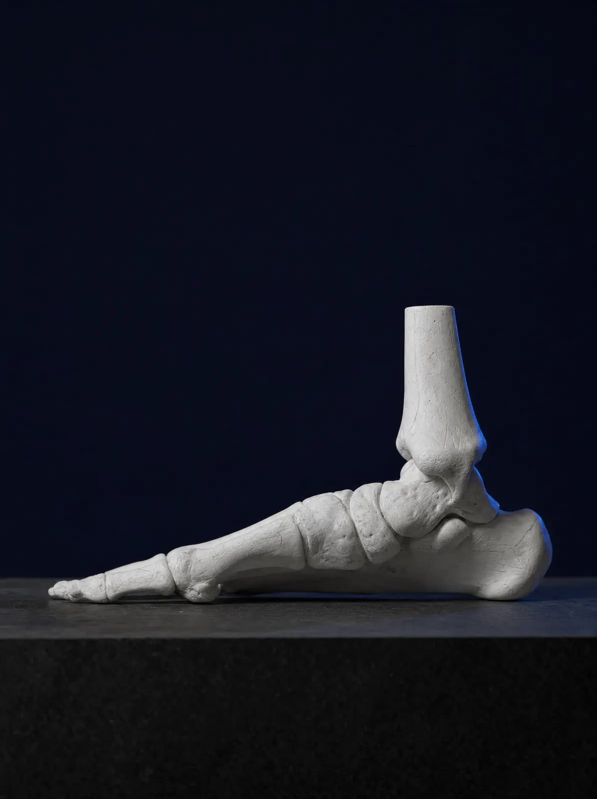
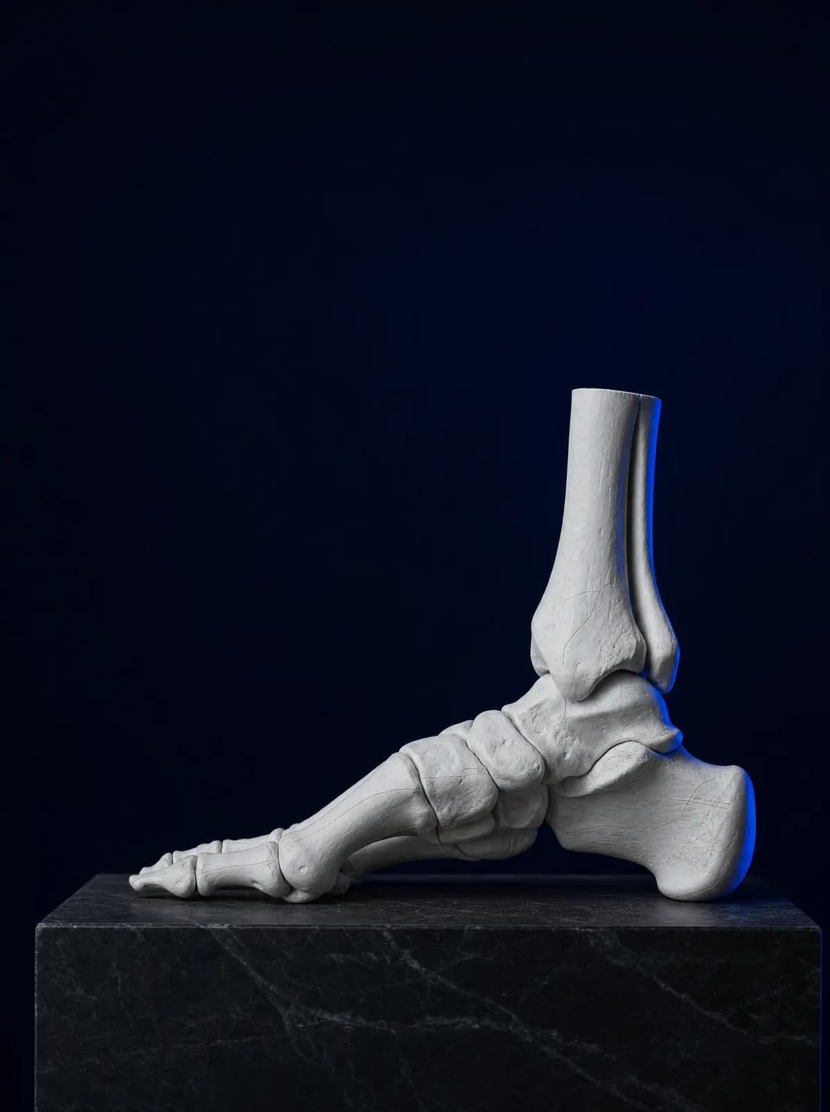
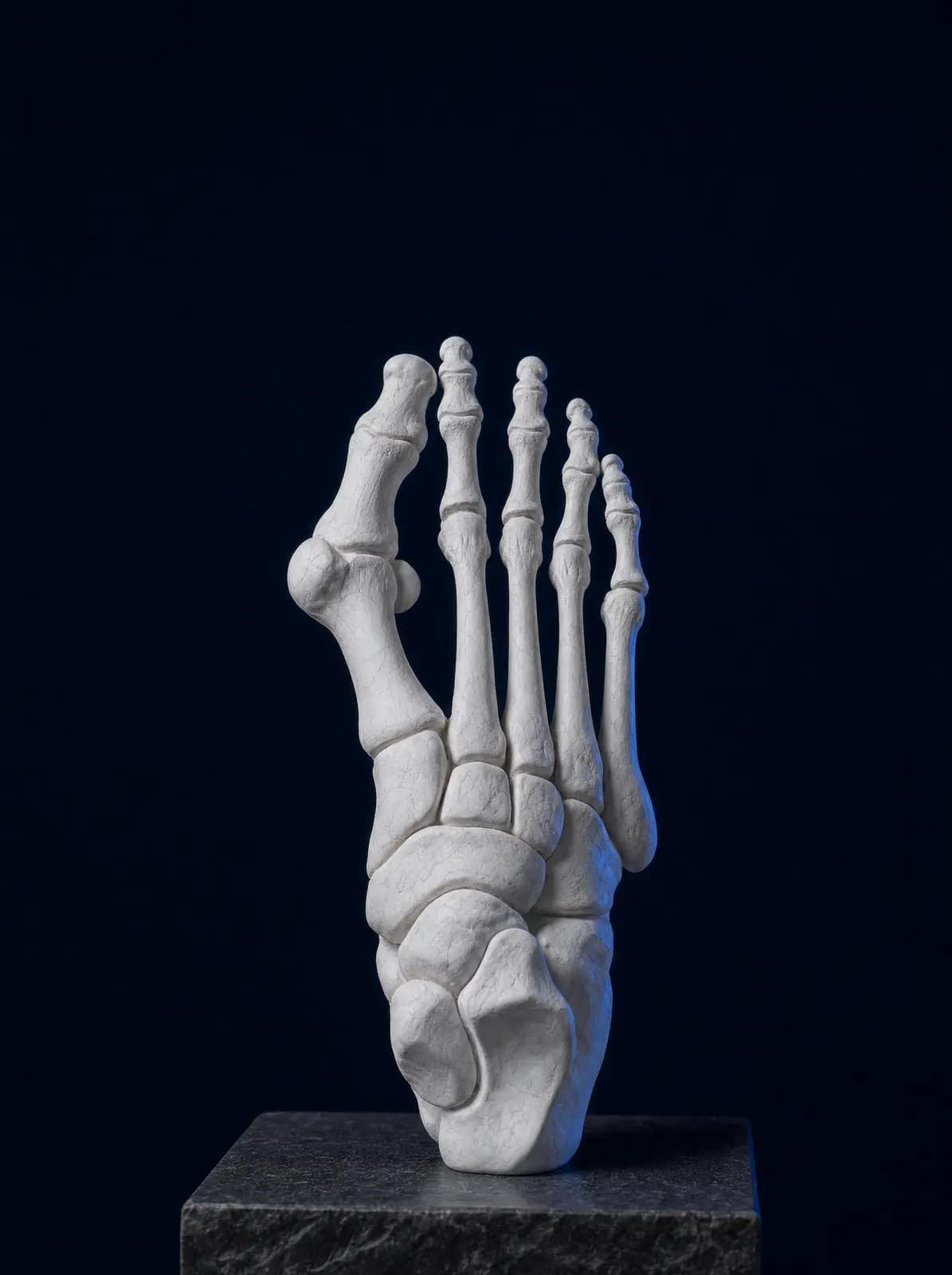
An arch rises along the inner edge of the foot between the heel and the bases of the toes. Supported by bone alignment, ligaments and tendons, particularly the tibialis posterior tendon on the inner side, it acts like a shock absorber that stretches and recoils during walking. Flat foot (pes planus) is marked lowering or complete loss of this arch when standing; the heel often tilts outwards.
Flat foot is a foot posture rather than a single disease. Distinguishing two forms guides treatment. In flexible flat foot, the arch reappears when the foot is off the ground or on tiptoe; the foot is supple and mobile. In rigid flat foot, no position restores the arch and movement is restricted; an underlying structural cause is investigated.
Flexible flat feet are common and usually cause no symptoms. Symptom-free flat feet require no treatment; pain and loss of function, rather than the X-ray appearance, guide decisions.
What Are the Symptoms?
When symptoms occur, the most common descriptions are:
- Pain in the sole and inner foot after prolonged walking, standing or sport
- Swelling and tenderness on the inner ankle
- Rapid wear on the inner edge of the shoe sole
- Fatigue and pain in the calf, knee and lower back
- Difficulty rising onto tiptoes or standing on one foot
- In children, avoiding long walks, asking to be carried and tiring quickly while running
Flat Feet in Children
Babies are born with flat feet. The arch develops gradually from the first years of walking and becomes more apparent towards school age in most children; in some it develops until adolescence. A flat-looking sole in a young child is not itself a disease. The fat pad under young children's feet also hides the arch, giving a flat appearance.
Families most often ask whether insoles are needed. There is no strong evidence that insoles permanently change the arch in children with painless, flexible flat feet; routine insoles are therefore not recommended for children without symptoms. Insoles may be used to relieve pain in children who have discomfort or complain after prolonged walking. Flexible-soled shoes that do not squeeze the feet are sufficient; walking barefoot on uneven surfaces such as grass or sand exercises the foot muscles.
Assessment focuses on warning signs: one-sided flat foot, pain, stiffness, a short Achilles tendon, significantly altered walking and worsening symptoms. If any are present, causes other than flexible flat feet are investigated. For other walking issues, such as intoeing and toe walking, see intoeing and gait disorders in children.
Adult Acquired Flatfoot
Painless flat feet present since childhood and newly developed flat feet in adulthood are different conditions. The most common cause of adult arch collapse is dysfunction of the tibialis posterior tendon, which passes along the inside of the ankle and supports the arch. Excess weight, prolonged standing at work, diabetes, rheumatic conditions and previous injuries increase the risk.
Early on, pain and swelling affect the inner ankle while the arch remains flexible. As the condition progresses, the heel shifts outwards, the forefoot turns out and the foot gradually stiffens; the ankle joint may also deteriorate in advanced stages. New adult flat foot and inner ankle pain should therefore not be ignored.
Treatment depends on the stage. In early disease, reducing load, using an arch-supporting insole or ankle brace, following a tendon-strengthening programme and managing pain are sufficient for most patients. If the foot has become stiff, the deformity has progressed and pain cannot be controlled with non-surgical measures, surgical options are discussed: tendon repair or transfer, corrective operations on the heel bone and, in advanced cases, fusion of selected joints. The decision depends on foot flexibility, stage, age and expectations.
How Is It Diagnosed?
Examination compares the arch while standing and with the foot unloaded, includes a tiptoe-rise test and assesses heel alignment from behind. Achilles tightness and foot joint range of movement are checked to distinguish flexible from rigid flat feet.
If symptoms are present, standing weight-bearing foot X-rays show bone alignment and joint condition. For a rigid foot, CT may identify bridging between bones, while MRI assesses tendons and ligaments. Imaging is generally unnecessary for painless flexible flat feet.
Rigid Flatfoot: Tarsal Coalition (Joined Bones)
Tarsal coalition is a connection between bones at the back of the foot, most commonly between the heel bone and ankle-region bones, through a bridge of bone, cartilage or fibrous tissue. It is present from birth, but symptoms usually begin in the second decade of life as the bridge stiffens. The typical pattern is an adolescent with recurrent ankle sprains, a rigid flat foot and increased pain after sport.
Restricted foot movement is identified on examination, and the diagnosis is confirmed by CT or MRI. First-line treatment is non-surgical: activity modification, arch-supporting insoles, a period of rest in a splint or cast, and pain management. If symptoms persist, surgical removal of the bridge or, in selected cases, corrective joint procedures are considered. This issue is assessed separately during consultation, and planning follows the imaging findings.
What Should You Consider in Daily Life?
Simple exercises for the sole and calf muscles, such as heel raises, gathering a towel with the toes and calf stretching, support the structures maintaining the arch. Weight management directly reduces foot loading. Cushioned, arch-supporting shoes can help people who stand for long periods.
Assessment is needed if foot pain persists for several weeks, flat foot develops on one side only, the foot becomes rigid or a child's gait deteriorates noticeably. You can book by telephone or WhatsApp at AC CLINIC in Altınordu, Ordu, for paediatric and adult foot assessment, planning of weight-bearing X-rays, and an insole and exercise programme.
Frequently Asked Questions
There is no strong evidence that insoles permanently alter the arch in children with painless flexible flat feet, so routine insoles are not recommended. They may be used for comfort if long walks cause pain and fatigue. The decision is made during assessment.
The arch develops gradually in the years after walking begins and becomes apparent by school age in most children; in some, development continues into adolescence. Even if an arch does not develop, treatment is unnecessary without pain or functional loss.
Surgery is discussed for pain persisting despite non-surgical treatment, progressive deformity and loss of function, not simply a flat-looking sole. It may also be considered for persistent symptoms from causes of rigid flat foot such as tarsal coalition.
Many people with painless flexible flat feet participate in sport and heavy physical work without problems. Official medical decisions are made by authorised bodies according to applicable regulations and examination findings; this page is for patient information only.
Repeated ankle sprains beginning in adolescence and a stiff foot with restricted movement may suggest tarsal coalition. Examination and imaging distinguish it; treatment starts with non-surgical approaches.
You can book by telephone or WhatsApp at AC CLINIC in Altınordu. Bring your child's most-worn shoes, or previous X-rays and MRI scans for an adult, to help the assessment.
References
Appointments and Contact
Call or contact us on WhatsApp to arrange an appointment. Briefly describe your symptoms; we try to schedule your examination and any necessary imaging on the same day.
- Opening hours
- 08:30–18:00Monday – Saturday
The clinic is not a 24/7 emergency department. For a fracture, dislocation, open wound, or inability to bear weight with fever, call 112 or attend the nearest hospital emergency department.